| Gastroenterology Research, ISSN 1918-2805 print, 1918-2813 online, Open Access |
| Article copyright, the authors; Journal compilation copyright, Gastroenterol Res and Elmer Press Inc |
| Journal website http://www.gastrores.org |
Case Report
Volume 12, Number 3, June 2019, pages 174-175
Tackling the Diagnosis: Solid Pseudopapillary Tumor of the Pancreas in a Young Man
Marilia Camposa, e, James Campbellb, Hernando Gonzalezc, Mufaddal Najmuddind
aDepartment of Medicine, Hennepin Healthcare, Minneapolis, MN, USA
bDepartment of Gastroenterology, University of Minnesota, Minneapolis, MN, USA
cDepartment of Gastroenterology, Hennepin Healthcare, Minneapolis, MN, USA
dDepartment of Pathology, University of Minnesota, Minneapolis, MN, USA
eCorresponding Author: Marilia Campos, Department of Medicine, Hennepin Healthcare, 701 Park Ave. (G5), Minneapolis, MN 55415, USA
Manuscript submitted March 2, 2019, accepted March 11, 2019
Short title: Solid Pseudopapillary Tumor of the Pancreas
doi: https://doi.org/10.14740/gr1170
| Abstract | ▴Top |
Solid pseudopapillary tumor (SPT) of the pancreas is an extremely rare pancreatic tumor. It usually goes undetectable for a prolonged period of time given its lack of clinical symptoms. But when detected, it has a malignancy potential of 15%. Here we present a case of SPT diagnosed incidentally in a 17-year-old man during a routine evaluation for an abdominal trauma.
Keywords: Solid pseudopapillary tumor; Pancreatic tumor; Young man
| Introduction | ▴Top |
Solid pseudopapillary tumor (SPT) of the pancreas represents only about 1% of all pancreatic tumors. It is more common in women (10:1) and has a low malignancy potential of approximately 15%. This case describes the incidental finding of an SPT in a healthy 17-year-old male patient.
| Case Report | ▴Top |
A 17-year-old otherwise healthy man presented for an abdominal trauma evaluation after being tackled at a football game. No major injuries were found.
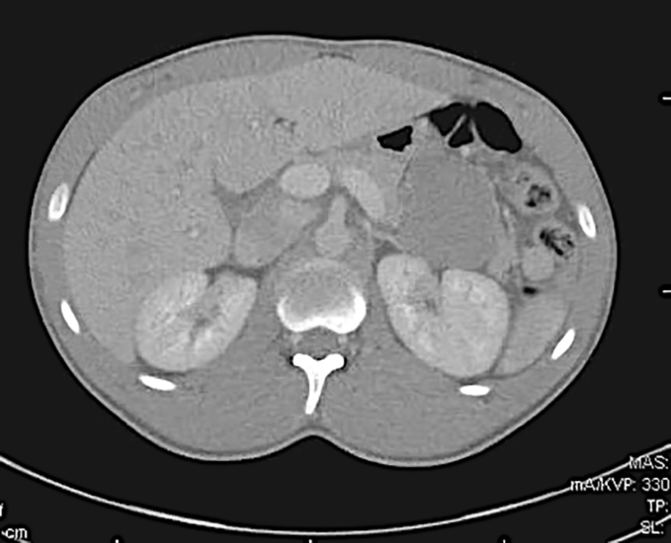
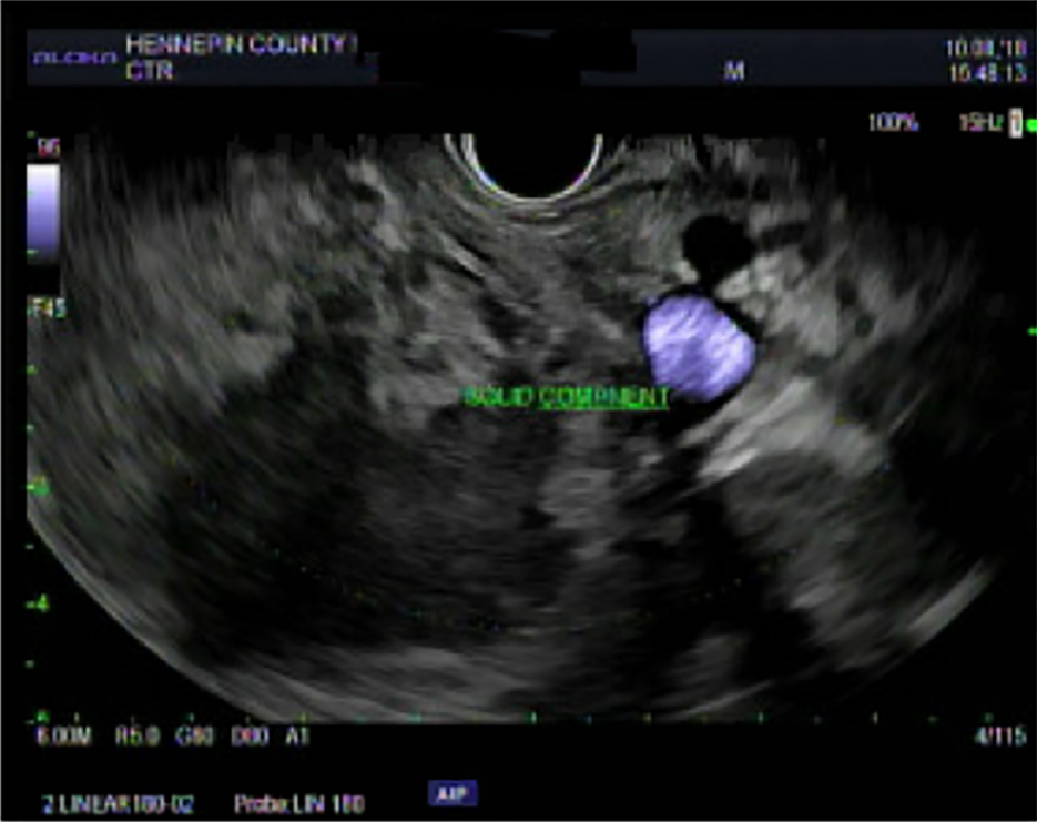
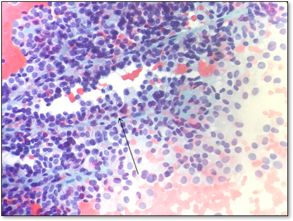
During the evaluation, an incidental 5.8-cm pancreatic mass was noted on contrast-enhanced abdominal computed tomography (CT) (Fig. 1). He denied any previous symptoms, including abdominal pain, mass and/or weight loss. Magnetic resonance imaging (MRI) revealed a predominantly cystic pancreatic tail mass with irregular walls. The patient underwent an endoscopic ultrasound (EUS) (Fig. 2) the following day, which showed a soft tissue mass with multiple cystic components. Fine-needle aspiration specimen revealed a solid pseudopapillary neoplasm (Fig. 3). The cytopathological (cytomorphologic) study showed small clusters and single cells, few clusters showing a papillary architecture with central fibrovascular core and loosely attached neoplastic cells. Immunostains performed on the cell block showed that tumor cells are positive for beta catenin, Sox-11, CD10, PR and vimentin. He was hospitalized for a total of three nights and had close follow-up in the GI clinic and surgery clinic. Tentative plan is for him to undergo pancreatic resection of the tumor.
 Click for large image | Figure 1. Contrast-enhanced computed tomography evidencing 5.8 cm pancreatic mass. |
 Click for large image | Figure 2. Endoscopic ultrasound (EUS) showing a soft tissue mass with multiple cystic areas. |
 Click for large image | Figure 3. Cytopathological (cytomorphologic) study showing small clusters and single cells, few clusters showing a papillary architecture with central fibrovascular core and loosely attached neoplastic cells. |
| Discussion | ▴Top |
SPT represents only about 1% of all pancreatic tumors and is very rare in young men as in the case presented. Most cases are found in young women (10:1) and the mean age of presentation is 22 years [1]. It is often clinically asymptomatic, as in our patient; however, it can also present with abdominal pain, a gradually enlarging mass, or rarely with jaundice [2]. Most SPTs are benign tumors; however, malignancy can occur in about 15% of cases, thus surgical resection is recommended [3, 4]. The most common metastatic sites are liver and omentum, and metastases are more commonly associated with tumors located in the pancreatic body and tail [3, 5]. SPTs rarely manifest themselves clinically and this case demonstrates how an incidental finding saved this young man’s life. The patient will now have an overall 5-year survival of 97% given he will undergo surgical resection [3].
Acknowledgments
None to declare.
Financial Disclosure
None to declare.
Conflict of Interest
None to declare.
Informed Consent
Informed consent was obtained from the patient.
Author Contributions
Marilia Campos: main author. James Campbell: reviewer. Hernando Gonzalez: reviewer. Mufaddal Najmuddin: provided with pathology images.
| References | ▴Top |
- Mergener K, Detweiler SE, Traverso LW. Solid pseudopapillary tumor of the pancreas: diagnosis by EUS-guided fine-needle aspiration. Endoscopy. 2003;35(12):1083-1084.
doi pubmed - Sun CD, Lee WJ, Choi JS, et al. Solid-pseudopapillary tumours of pancreas: 14 years' experience. ANZ J Surg. 2005;75(8):664-689.
doi - Chen X, Zhou GW, Zhou HJ, Peng CH, Li HW. Diagnosis and treatment of solid-pseudopapillary tumors of the pancreas. Hepatobiliary Pancreat Dis Int. 2005;4(3):456-459.
- Mulkeen AL, Yoo PS, Cha C. Less common neoplasms of the pancreas. World J Gastroenterol. 2006;12(20):3180-3185.
doi pubmed - Kim MJ, Choi DW, Choi SH, Heo JS, Sung JY. Surgical treatment of solid pseudopapillary neoplasms of the pancreas and risk factors for malignancy. Br J Surg. 2014;101(10):1266-1271.
doi pubmed
This article is distributed under the terms of the Creative Commons Attribution Non-Commercial 4.0 International License, which permits unrestricted non-commercial use, distribution, and reproduction in any medium, provided the original work is properly cited.
Gastroenterology Research is published by Elmer Press Inc.


