

| Gastroenterology Research, ISSN 1918-2805 print, 1918-2813 online, Open Access |
| Article copyright, the authors; Journal compilation copyright, Gastroenterol Res and Elmer Press Inc |
| Journal website https://www.gastrores.org |
Case Report
Volume 15, Number 5, October 2022, pages 278-283

Adult Intussusception in Chronic Marijuana Users
Jiten P. Kothadiaa, b, e ![]() , Anwesh Dashc, Rajanshu Vermac
, Anwesh Dashc, Rajanshu Vermac ![]() , Kyle Kreitmanc, Peter D. Snellc, Mohammad K. Ismaild
, Kyle Kreitmanc, Peter D. Snellc, Mohammad K. Ismaild
aJames D. Eason Transplant Institute, Methodist University Hospital, Memphis, TN, USA
bDivision of Transplant Surgery, Department of Surgery, University of Tennessee Health Science Center, Memphis, TN, USA
cDivision of Gastroenterology, Department of Medicine, University of Tennessee Health Science Center, Memphis, TN, USA
dDepartment of Medicine, Division of Gastroenterology, Washington University School of Medicine, St. Louis, MO, USA
eCorresponding Author: Jiten P. Kothadia, James D. Eason Transplant Institute, Methodist University Hospital, Memphis, TN 38104, USA
Manuscript submitted July 7, 2022, accepted August 12, 2022, published online October 19, 2022
Short title: Adult Intussusception in Chronic Marijuana Users
doi: https://doi.org/10.14740/gr1554
| Abstract | ▴Top |
Intussusception is common in children, but it is rare in adults. The most common causes of adult intussusception (AI) are due to a pathological lead point with a common etiology being malignancy. Intra-luminal irritants should be considered the possible etiology of intussusception in patients without a pathological lead point. Marijuana use has increased dramatically in the United States over the last decade. With increasing public acceptance and legalization of marijuana, various adverse side effects have become more prominent. Marijuana has been shown to disrupt gastrointestinal tract motility by inhibiting cholinergic mechanisms. Here we describe four cases of AI who are chronic marijuana users. This well-referenced review gives attention to the harmful effects of marijuana, given the increasing use of marijuana and its derivatives in the United States.
Keywords: Intussusception; Abdominal pain; Chronic marijuana use; Bowel obstruction
| Introduction | ▴Top |
Intussusception of the bowel refers to the invagination of a proximal segment (intussusceptum) into an adjacent distal segment (intussuscipiens). It is common in the pediatric population but rare in adults, with an annual incidence of 2 - 3 cases per 1,000,000 population [1]. AI represents only 1-5% of mechanical small bowel obstructions in adults [1, 2]. It can be classified as primary or secondary based on etiology. The most cases of intussusception in children are due to primary and benign, while almost 90% cases of AI are secondary to pathology that acts as a lead point. The secondary causes of intussusception include neoplasms, Meckel’s diverticulum, strictures, endometriosis, polyps, post-operative adhesions, and arteriovenous malformations. Neoplasms are the most frequent cause of secondary AI representing up to 63% of all adult cases [2, 3].
The diagnosis of AI is challenging due to non-specific symptoms and requires a high index of clinical suspicion. The common symptoms include crampy abdominal pain, nausea and vomiting. Furthermore, intussusception can cause intestinal edema secondary to venous, and lymphatic congestion from mesentery entrapment in the bowel. If not resolved, this obstruction can eventually lead to vascular compromise and increase the risk of perforation and patient may present with bloody diarrhea and signs of peritonitis. The detailed history, physical exam, and laboratory studies can aid in diagnosis process, but imaging such as computed tomography (CT) of the abdomen is usually needed to confirm the diagnosis.
The cannabinoids have anti-inflammatory effects and have shown a beneficial role in treating chemotherapy-related nausea and vomiting, multiple sclerosis (MS)-related spasticity and chronic pain [4]. The studies have shown anti-peristaltic effects of cannabinoids through activation of cannabinoid receptor type 1 (CB1) receptors involving epithelial cells of the gastrointestinal (GI) tract and myenteric and submucosal nerve plexus [7]. There are several reported cases of AI in association with chronic marijuana use in the literature [1, 5-9]. We present four patients with history of chronic marijuana use who presented with intussusception.
| Case Reports | ▴Top |
Case 1
A 27-year-old male with a past medical history of gastroesophageal reflux disease and peptic ulcer disease presented with severe abdominal pain for 3 days. The patient did not come to the emergency department immediately because he assumed the pain was a flare-up of peptic ulcer disease. He had multiple prior esophagogastroduodenoscopies (EGDs) that showed gastritis, pre-pyloric ulcers, and duodenitis. He had also been treated for H. pylori with bismuth-based quadruple therapy. The pain was sharp, 7/10 in severity, and localized to the epigastric region without radiation. The patient had associated nausea with multiple episodes of emesis. The patient denied dysphagia, diarrhea, melena, or hematochezia. The patient denies alcohol use but endorses marijuana use up to 2 - 3 g per day for 10 years and cigarette use up to 1 pack per day for 6 years.
On physical examination, he had localized severe epigastric tenderness and guarding. Vitals were within normal limits. Abnormal lab results included an elevated red blood cell (RBC) count at 6.54, low mean corpuscular volume at 74.8 (80 - 100), and high red cell distribution width at 18.2. White blood cell (WBC) count was normal. CT scan revealed small bowel intussusception (SBI) with no evidence of inflammation in the colon or appendix (Fig. 1). The patient was emergently taken to surgery for midline laparotomy. After entering the abdomen, an area of dilated small bowel with proximal and distal collapse was seen in the mid-jejunum. This section of the jejunum was carefully straightened, leading to improvement in the distension. The segment of the small bowel was carefully palpated for signs of intraluminal lesions. The small bowel was examined from the ligament of Treitz to the terminal ileum, and no abnormality could be discovered. No evidence of tumors or masses was apparent. The patient was discharged home in 2 days upon clinical improvement.
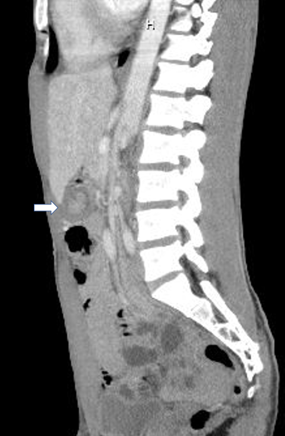 Click for large image | Figure 1. Computed tomography (CT) scan (sagittal with arrow) showing telescoping of small bowel within itself consistent with small bowel intussusception with no evidence of inflammation in the colon or appendix. |
Case 2
A 33-year-old female with a past medical history significant for alcohol abuse, hypertension, gallstone pancreatitis, and esophagitis presented with a 1-day history of abdominal pain, nausea, vomiting, and diarrhea. The pain was cramping in nature, periumbilical in location, and worsened over 24 h. The patient denies any episodes of hematemesis, hematochezia, or melena. She recalled previous episodes of mild cramping abdominal pain but none that matched the severity of the current episode. She reported daily marijuana use for the past 10 years.
On admission, she was afebrile, with a respiratory rate of 16/min and a heart rate of 115 beats/min with 146/88. The abdomen was soft, non-distended, and tender around the umbilicus, with no peritoneal signs. Initial laboratory work was unremarkable, including complete blood count, serum chemistry, liver test, urinalysis, and a urine pregnancy test. CT scan of the abdomen with contrast showed fatty liver, normal spleen and distal small bowel, and terminal ileum intussusception into the colon (Fig. 2). Subsequent CT enterography showed nonspecific enteritis and resolution of intussusception without perforation or obstruction. Given CT enterography findings, no surgical intervention was planned, and she was managed conservatively. Since no other cause for intussusception could be identified, she was counseled about substance use cessation. The patient subsequently underwent a colonoscopy, which showed mild diverticulosis without bleeding and a small polyp in the descending colon. The patient improved with expectant management and was discharged on day 3 with outpatient follow-up.
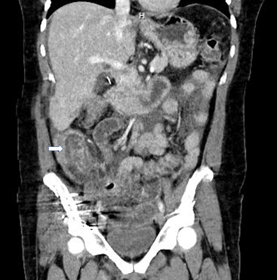 Click for large image | Figure 2. Computed tomography (CT) scan (coronal with arrow) of abdomen showing terminal ileum intussusception into the colon. |
Case 3
A 29-year-old male with no significant past medical history presented with 2 weeks of diffuse crampy abdominal pain, nausea, vomiting, and diarrhea. The patient endorsed occasional hematochezia and reduced oral intake but noted no hematemesis, melena, or constipation. The patient reported daily marijuana use of approximately 1 g for the past 8 years but denied alcohol, tobacco, or illicit drug use. He had no personal or family history of GI disease.
On admission, he was hemodynamically stable and in no apparent distress. A physical examination showed a soft, non-distended, mildly tender abdomen with no peritoneal signs. Labs were significant for an elevated WBC count at 13.56 × 10³/µL (4.2 - 10.2), and elevated lipase at 74.8 U/L (10 - 70) with normal liver tests. CT scan of the abdomen with contrast showed jejuno-jejunal intussusception in the left mid-abdomen with sigmoid and descending colon wall thickening concerning active inflammation (Fig. 3). Infectious workup was unremarkable. The patient was treated conservatively with intravenous (IV) fluids and antiemetics for nausea. The surgery team was consulted for evaluation, but no surgical intervention was necessary as the patient’s intussusception resolved with conservative management. The patient was discharged home on day 2 with outpatient follow-up.
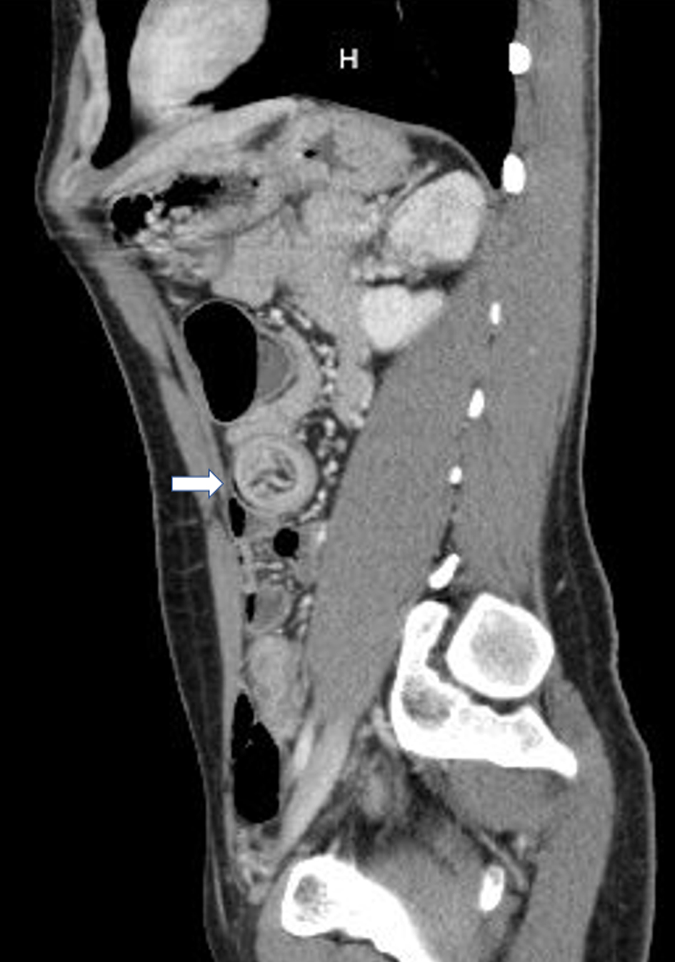 Click for large image | Figure 3. Computed tomography (CT) scan (sagittal with arrow) showing jejuno-jejunal intussusception in the left mid-abdomen with colon wall thickening. |
Case 4
A 36-year-old Caucasian male came to the emergency room with a chief complaint of abdominal pain, nausea, vomiting, and abdominal pain for 1 week. He reported abdominal pain mainly in the lower abdomen 5/10 in severity, cramping in nature, non-radiating with no aggravating or relieving factor. Reported vomitus was non-bilious and non-bloody. He denies any weight loss or any upper-lower GI bleeding. He reported daily marijuana use for the past 15 years but denied alcohol, tobacco, or illicit drug use. No recent travel history was noted.
On admission, his vitals were stable, and on the exam, he was noted to have mild generalized tenderness on the lower quadrant without any guarding, rigidity, or peritoneal signs. Laboratory studies showed complete blood cell count, comprehensive metabolic profile, liver chemistries, normal liver chemistry, and amylase and lipase. The patient underwent a CT of abdomen with contrast that showed multiple small bowel short segment intussusception areas without proximal dilatation (Fig. 4). Infectious workup was unremarkable. General surgery and gastroenterology were consulted for further evaluation. He was treated with conservative management with bowel rest, IV fluids, and anti-emetics. Subsequently, the next day, the patient clinically improved the complete resolution of symptoms and had bowel movements and had a small bowel follow-through series that came unremarkable. He underwent EGD that showed erosive gastritis, clean base multiple ulcers antrum (3 - 5 mm), and duodenal erythema. The patient was discharged on day 2 upon clinical improvement.
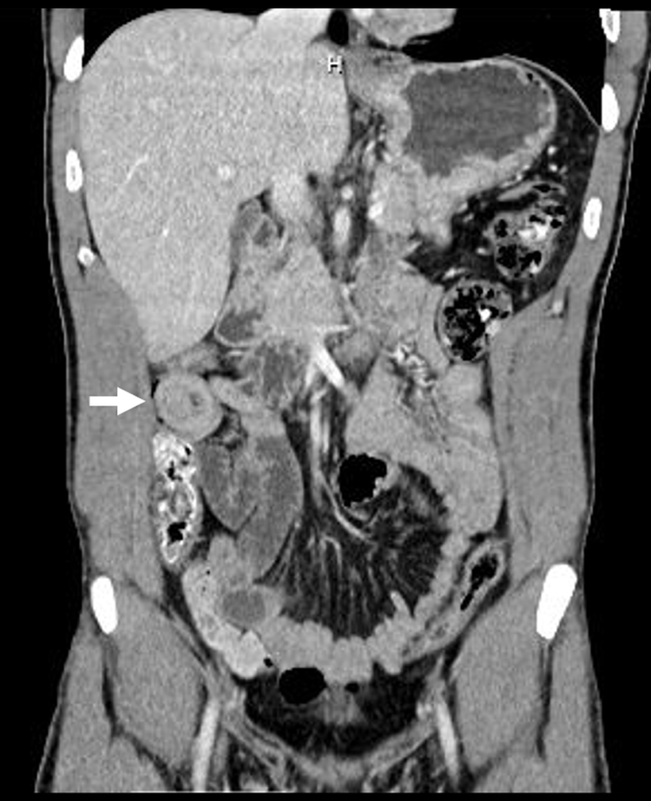 Click for large image | Figure 4. Computed tomography (CT) of abdomen (coronal with arrow) showing small bowel short segment intussusception areas without proximal dilatation. |
| Discussion | ▴Top |
Marijuana products are the most universally used recreational drug across the world [10]. In the United States, 17.9% (around 49.6 million) of people aged 12 or older reported using marijuana over the past 12 months [11]. Trends indicate marijuana use has increased markedly among younger populations, with a higher percentage of high school students using marijuana products. The gender gap in marijuana use has also widened with more men abusing the drug than women [12]. With such increasing usage of marijuana, learning about the adverse effects of the drug, especially its effect on the GI system, has become more important than ever before.
Tetrahydrocannabinol (THC), the primary active ingredient of marijuana products, acts through many receptors in the GI tract. The primary effects are through the CB receptor system. CB1 receptors are located in the myenteric and submucosal neurons of the GI tract, while CB2 receptors are located primarily on inflammatory and epithelial cells [13]. Activation of these receptors results in inhibition of acetylcholine release in pre-synaptic neurons leading to slowed contractile activity and peristalsis. Studies have also shown delayed gastric emptying and inhibition of postprandial colonic tone from chronic marijuana use [13]. This dissonance in peristalsis from chronic marijuana use has been postulated to provide the lead point for intussusception.
The diagnosis of AI is challenging due to non-specific presenting symptoms, and suspecting marijuana use as the cause of AI requires a thorough history and a very high index of clinical suspicion. As more than 90% of AI cases are due to secondary causes, it is crucial that patients undergo appropriate testing to diagnose etiology. Abdominal radiography is often first ordered to evaluate bowel obstruction, but it has low sensitivity and specificity for diagnosis. Abdominal CT is the most sensitive and diagnostic test of choice that shows a “target” sign or “sausage shaped” soft-tissue mass pathognomonic of intussusception [14-16]. Endoscopy is often used adjunctively to confirm the location of intussusception and visualize the lead point.
Management of AI depends on the severity of the clinical presentation. Reducing the bowel using barium or air enema employed in pediatrics is often inadequate treatment for adults due to the theoretically higher likelihood of spreading malignancy and perforation [17, 18]. Historically, surgical intervention with explorative laparotomy or laparoscopy remains the mainstay form of treatment of AI. It allows evaluation of areas of ischemic bowel and the resection of the lead point since malignancy comprises up to 50% of cases [19]. However, recent evidence favors preoperative or intraoperative reduction as first-line treatment and resection is warranted for in cases with vascular compromise or perforation [20].
Fernandez-Atutxa et al [9] first reported detailed a case series of chronic marijuana users who presented with intussusception. Since then, multiple published cases have supported a possible link between marijuana usage and AI [1, 5-8]. Causation between chronic marijuana use and intussusception has not been demonstrated, so most cases are identified after excluding other potential etiologies. No other etiology was identified in our patients, supporting chronic marijuana use as a possible cause of intussusception.
On review of the literature, 18 cases of AI in chronic marijuana users have been reported (Table 1) [1, 5-9]. All patients presented with non-specific symptoms like abdominal pain, nausea, vomiting, and diarrhea. The median age at presentation was 26 (interquartile range: 23 - 30), with no significant difference in incidence among males and females. The most common location of the intussusception was the jejunum, with one case involving the colon. Most patients were diagnosed with clinical suspicion and contrasted CT of abdominal scans. Expectant management with IV fluids, electrolyte replacement, and pain management was the cornerstone of treatment in all cases. Most cases spontaneously resolved, except for four patients who underwent diagnostic laparoscopy. Of these four cases, two patients required manual reduction while others had spontaneous resolution with negative laparoscopic findings. No obvious lead point was identified in any of these cases. In our case series, case 1 underwent laparotomy with manual reduction, while the rest of the patients had spontaneous resolution with conservative management. These cases highlight the importance of conservative management, and surgery should be reserved for those with failed conservative management or with acute abdomen.
 Click to view | Table 1. Reported Cases of Adult Intussusception in Chronic Marijuana Users |
Marijuana-induced AI continues to be a diagnosis of exclusion. AI is rare and has a high chance of having a malignant lead point, so patients should all undergo appropriate screening and imaging to rule out neoplastic sources. In a study by Wang et al [21], four out of 41 (10%) patients had “idiopathic” etiology where no pathological lead point was identified. This highlights the importance of a thorough search for the etiology of AI and detailed history taking. Our cases stress the importance of possible adverse effects of marijuana with increased legalization and therapeutic use of marijuana and its derivatives. Physicians must obtain detailed social history to identify marijuana use as a potential cause of idiopathic intussusception in adults.
Conclusion
Our cases depict the possible association between chronic marijuana and AI. Although rare, AI in the absence of pathological lead point, should alert physicians to look for intra-luminal irritants as a cause of intussusception. From a public health perspective, physicians should warn patients about the possible association between marijuana use and the development of intussusception. Further studies are needed to investigate the association between chronic marijuana use and AI.
Acknowledgments
None to declare.
Financial Disclosure
None to declare.
Conflict of Interest
The authors indicated no potential conflict of interest.
Informed Consent
Informed consent was obtained from the patients for publication of this manuscript.
Author Contributions
Jiten Kothadia, MD and Anwesh Dash, MD were involved in drafting the case report and the acquisition of available literature and revision of the manuscript. Rajanshu Verma, MD and Kyle Kreitman, MD reviewed manuscript. Mohammad K. Ismail, MD was involved in critical revision of the manuscript for important intellectual content.
Data Availability
The authors declare that the data supporting the findings of this study are presented within the content of this article.
| References | ▴Top |
- Zaidi SR, Khan ZH, Mukhtar K, Ahmed MM, Syed SH. A case of intussusception in a patient with marijuana use: coincidence or possible correlation? Cureus. 2020;12(3):e7493.
doi - Marinis A, Yiallourou A, Samanides L, Dafnios N, Anastasopoulos G, Vassiliou I, Theodosopoulos T. Intussusception of the bowel in adults: a review. World J Gastroenterol. 2009;15(4):407-411.
doi pubmed - Honjo H, Mike M, Kusanagi H, Kano N. Adult intussusception: a retrospective review. World J Surg. 2015;39(1):134-138.
doi pubmed - Weinstein J, et al. National Academies of Sciences, Engineering, and Medicine; Health and Medicine Division; Board on Population Health and Public Health Practice; Committee on Community-Based Solutions to Promote Health Equity in the United States. 2017.
- Khan ZH, et al. Intussusception in a patient with marijuana use: coincidence or possible correlation? American Journal of Gastroenterology. 2019. Lippincott Williams & Wilkins Two Commerce SQ, 2001 Market ST, Philadelphia.
- Silva V, Khalil K, Zaidi SR, Highsmith S, Tucker JI. Intussusception and chronic marijuana use in a young adult. Am J Case Rep. 2021;22:e932479.
doi - Kakish D, Alaoudi M, Welch B, Fan D, Meghpara M, Mandava N, Kumthekar N. Small bowel intussusception in marijuana users. J Surg Case Rep. 2020;2020(9):rjaa335.
doi pubmed - Prokopchuk O, Neumann PA, Huser N, Friess H, Wilhelm D. Multiple intestinal intussusceptions caused by highly impaired gastrointestinal motility in a patient with chronic cannabis consumption. J Surg Case Rep. 2019;2019(5):rjz160.
doi pubmed - Fernandez-Atutxa A, de Castro L, Arevalo-Senra JA, Langara E, Cabriada-Nuno JL. Cannabis intake and intussusception: an accidental association? Rev Esp Enferm Dig. 2017;109(2):157-159.
doi pubmed - Cohen K, Weinstein AM. Synthetic and Non-synthetic Cannabinoid Drugs and Their Adverse Effects-A Review From Public Health Prospective. Front Public Health. 2018;6:162.
doi pubmed - Abuse S. Key substance use and mental health indicators in the United States: Results from the 2020 National Survey on Drug Use and Health. 2021.
- Carliner H, Mauro PM, Brown QL, Shmulewitz D, Rahim-Juwel R, Sarvet AL, Wall MM, et al. The widening gender gap in marijuana use prevalence in the U.S. during a period of economic change, 2002-2014. Drug Alcohol Depend. 2017;170:51-58.
doi pubmed - Camilleri M. Cannabinoids and gastrointestinal motility: Pharmacology, clinical effects, and potential therapeutics in humans. Neurogastroenterol Motil. 2018;30(9):e13370.
doi pubmed - Gayer G, Apter S, Hofmann C, Nass S, Amitai M, Zissin R, Hertz M. Intussusception in adults: CT diagnosis. Clin Radiol. 1998;53(1):53-57.
doi - Gayer G, Hertz M, Zissin R. CT findings of intussusception in adults. Semin Ultrasound CT MR. 2003;24(5):377-386.
doi - Takeuchi K, Tsuzuki Y, Ando T, Sekihara M, Hara T, Kori T, Kuwano H. The diagnosis and treatment of adult intussusception. J Clin Gastroenterol. 2003;36(1):18-21.
doi pubmed - Begos DG, Sandor A, Modlin IM. The diagnosis and management of adult intussusception. Am J Surg. 1997;173(2):88-94.
doi - Eisen LK, Cunningham JD, Aufses AH, Jr. Intussusception in adults: institutional review. J Am Coll Surg. 1999;188(4):390-395.
doi - Weilbaecher D, Bolin JA, Hearn D, Ogden W, 2nd. Intussusception in adults. Review of 160 cases. Am J Surg. 1971;121(5):531-535.
doi - Sarr MG, Nagorney DM, McIlrath DC. Postoperative intussusception in the adult: a previously unrecognized entity? Arch Surg. 1981;116(2):144-148.
doi pubmed - Wang N, Cui XY, Liu Y, Long J, Xu YH, Guo RX, Guo KJ. Adult intussusception: a retrospective review of 41 cases. World J Gastroenterol. 2009;15(26):3303-3308.
doi pubmed
This article is distributed under the terms of the Creative Commons Attribution Non-Commercial 4.0 International License, which permits unrestricted non-commercial use, distribution, and reproduction in any medium, provided the original work is properly cited.
Gastroenterology Research is published by Elmer Press Inc.