Gastroenterology Research, ISSN 1918-2805 print, 1918-2813 online, Open Access
Article copyright, the authors; Journal compilation copyright, Gastroenterol Res and Elmer Press Inc
Journal website http://www.gastrores.org

Case Report

Volume 11, Number 1, February 2018, pages 79-82


Histologic Characterization of Kratom Use-Associated Liver Injury

Figure

Figure 1.
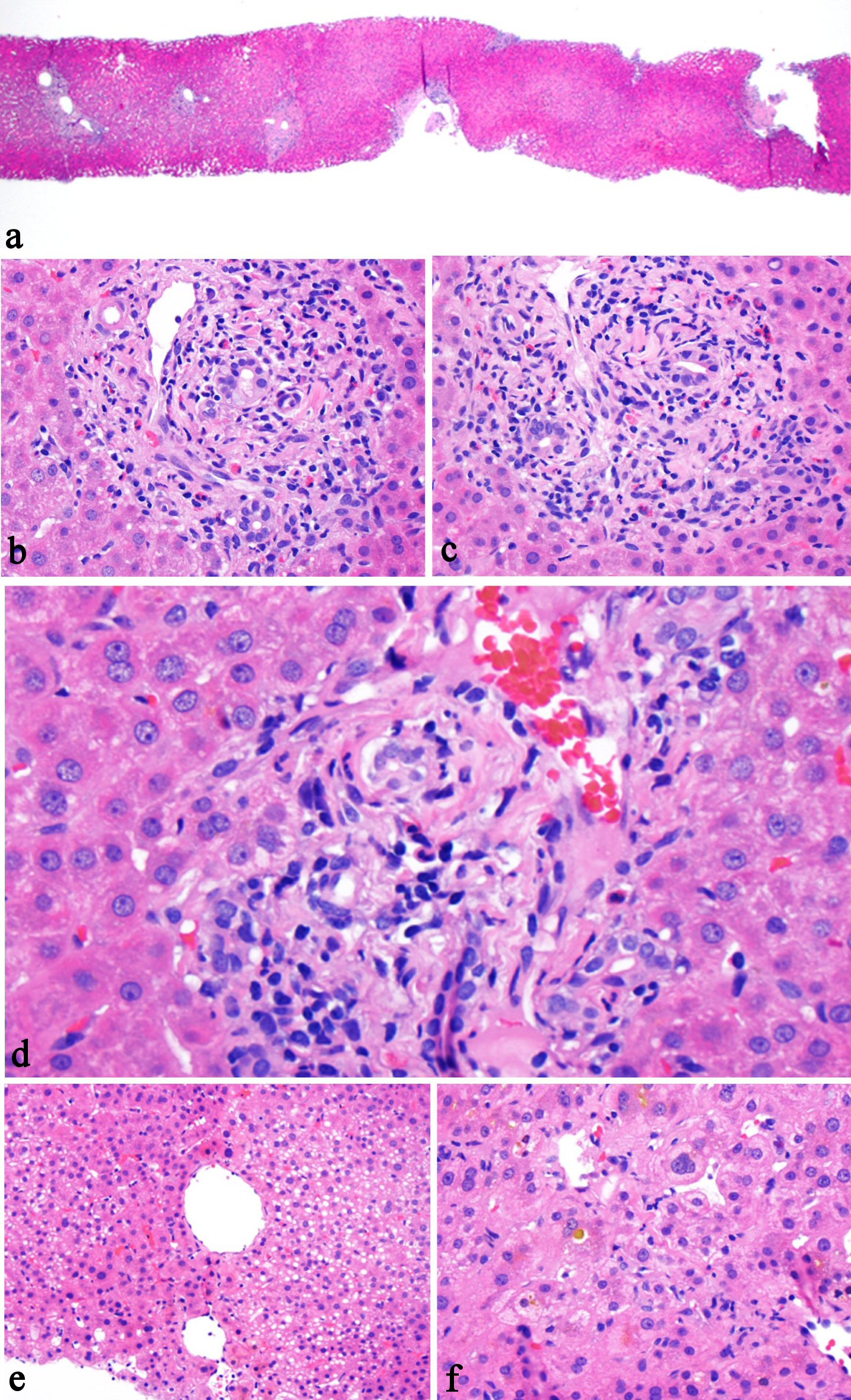
Figure 1. Histologic features of kratom-associated liver injury. The liver biopsy shows preserved normal lobular architecture (a, H&E stain, 20×), mixed portal inflammation composed of lymphocytes, eosinophils, rare plasma cells, and rare neutrophils (b, H&E stain, 200×), bile duct epithelial apoptosis (c, H&E stain, 200×), lymphocytic inflammation involves bile duct and ductules (d, H&E stain, 400×), focal steatosis (e, H&E stain, 100×), and zone 3 intrahepatocellular and canalicular cholestasis (f, H&E stain, 200×).

Table

Table 1. Laboratory Values at the Time of Presentation and After Cessation of Kratom
 
ALT (U/L)AST (U/L)Bilirubin (mg/dL)ALP (U/L)GGT (U/L)Comment
July16, 20163892205.1304Not doneKratom stopped
July 18, 20163601665.6266394
July 24, 20164101421.6266241
Normal values0 - 410 - 370.0 - 1.035 - 1295 - 61