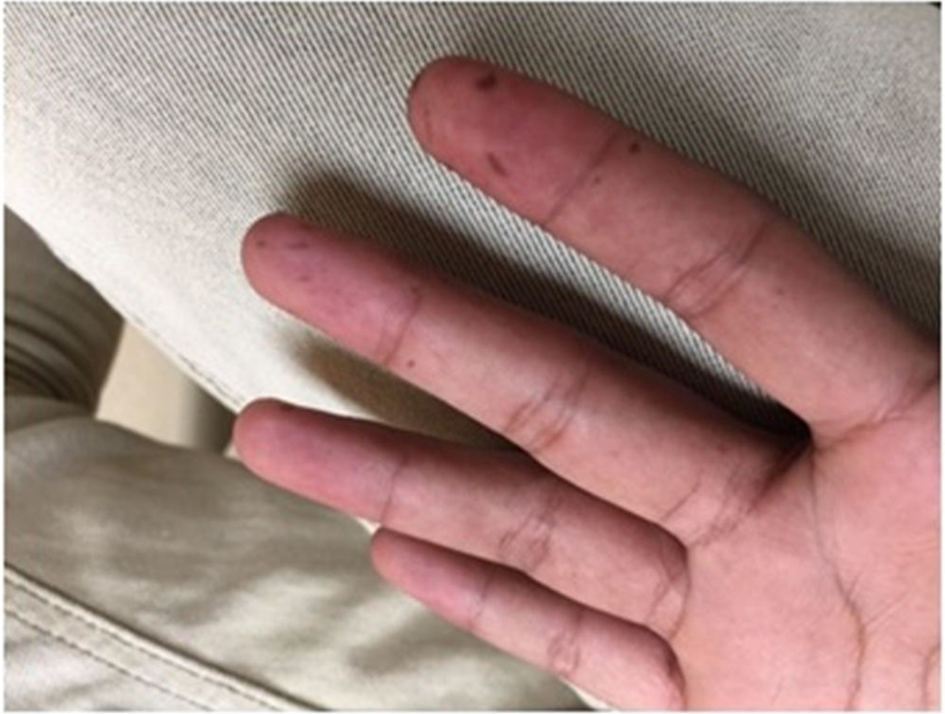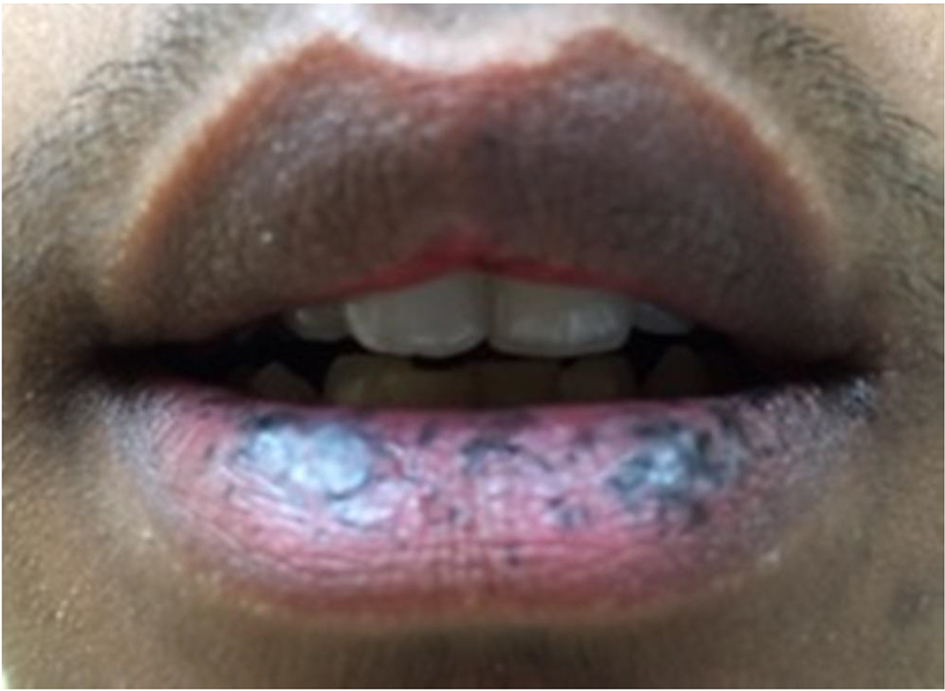
Figure 1. Characteristic hyperpigmented macules of Peutz-Jeghers syndrome on the patient’s lips.
| Gastroenterology Research, ISSN 1918-2805 print, 1918-2813 online, Open Access |
| Article copyright, the authors; Journal compilation copyright, Gastroenterol Res and Elmer Press Inc |
| Journal website http://www.gastrores.org |
Case Report
Volume 11, Number 2, April 2018, pages 150-153
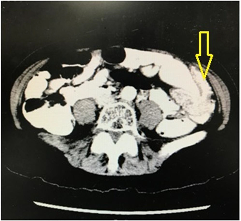
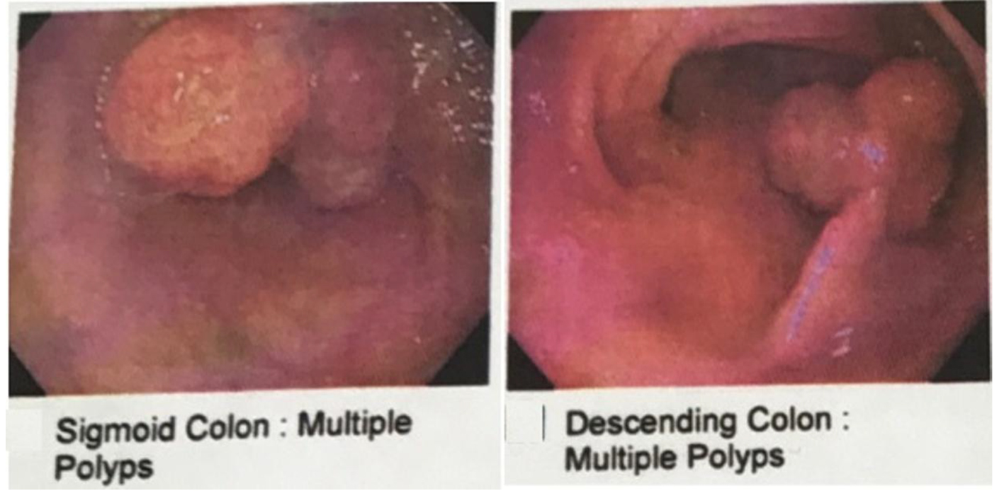
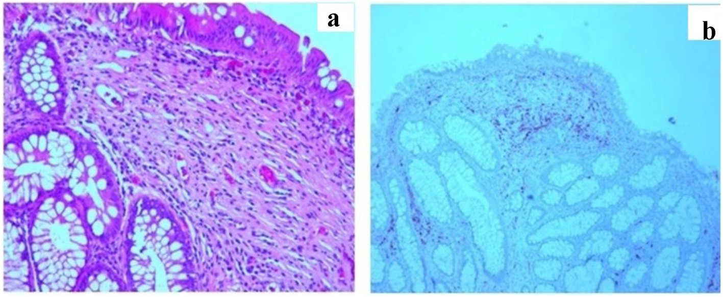
Peutz-Jeghers Syndrome Presenting as Colonic Intussusception: A Rare Entity
Figures