Figures
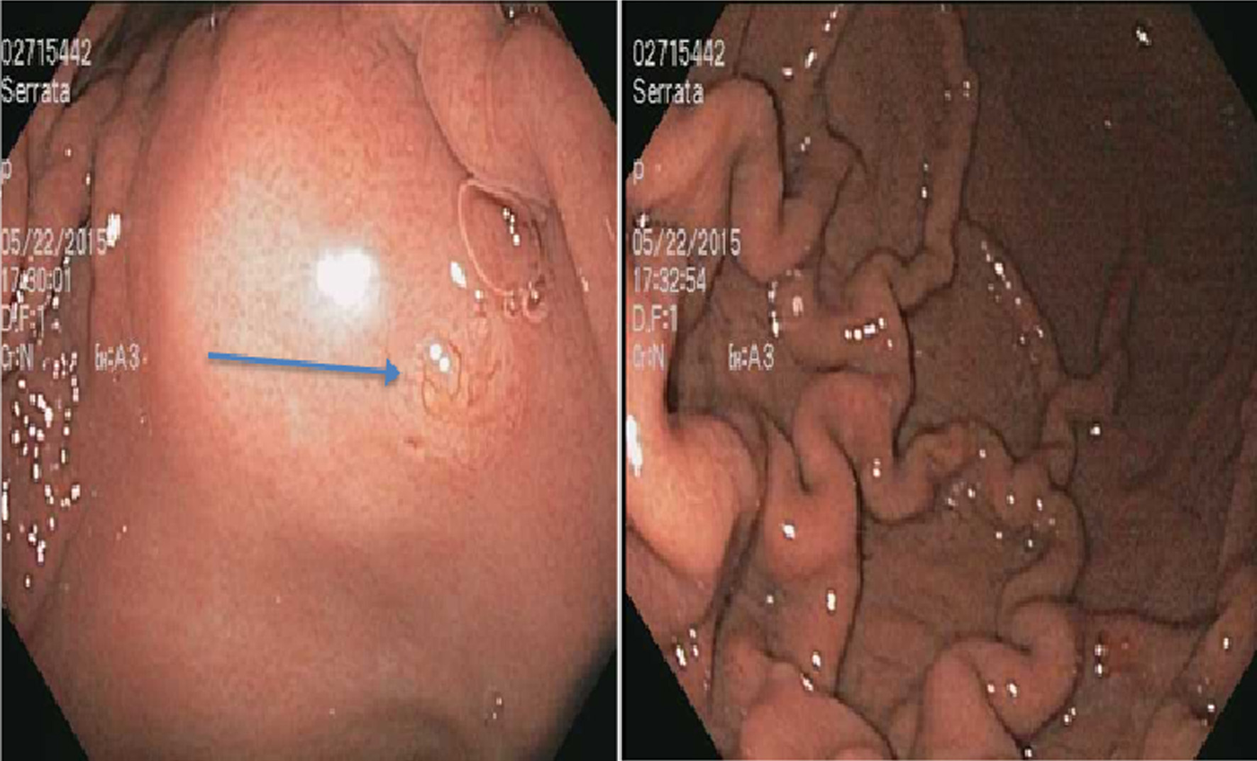
Figure 1. A 3 mm nodule on the lesser curvature of the stomach (arrow) (left) and prominent gastric folds (right).
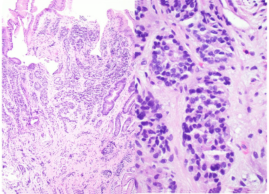
Figure 2. A well-differentiated neuroendocrine tumor in lamina propria with focal extension into muscularis mucosa consistent with a gastric carcinoid (hematoxylin and eosin stain, magnification: left × 40, right × 100).
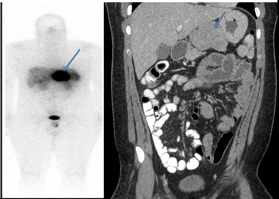
Figure 3. Octreotide scan showing abnormal uptake in the stomach consistent with a large gastric carcinoid without any evidence of metastatic disease (left). A computed tomography (CT) scan of abdomen and pelvis with contrast showed a lobulated soft tissue mass in the mid upper abdomen inseparable from inferior part of the liver and lesser curvature of the stomach (right).
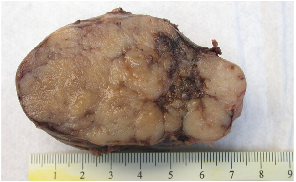
Figure 4. Macroscopic picture of peripancreatic mass with a lobulated cut surface. The mass was adherent to the lesser curve of stomach and anterior surface of pancreas.
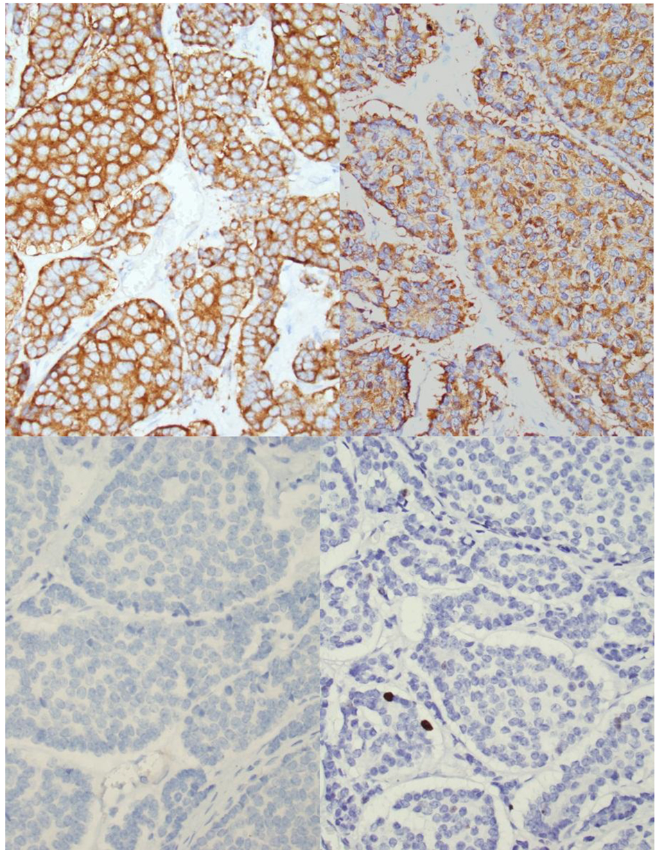
Figure 5. Histology of peripancreatic gastrinoma. Upper left (hematoxylin and eosin stain, please provide magnification). Immunohistochemistry revealed that the tumor cells are positive for chromogranin (upper mid), synaptophysin upper right) and gastrin (lower right) but negative for insulin (lower mid). Immunostain showed < 2% of tumor cells to be positive for Ki-67 (lower right).