Gastroenterology Research, ISSN 1918-2805 print, 1918-2813 online, Open Access
Article copyright, the authors; Journal compilation copyright, Gastroenterol Res and Elmer Press Inc
Journal website http://www.gastrores.org

Original Article

Volume 11, Number 1, February 2018, pages 25-30


Metastatic Renal Cell Carcinoma as Solitary Subcentimeter Polypoid Gastric Mucosal Lesions: Clinicopathologic Analysis of Five Cases

Figures

Figure 1.
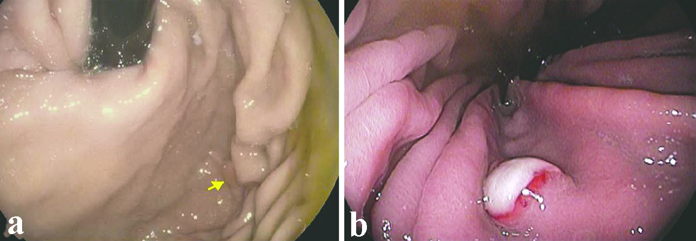
Figure 1. Representative endoscopic appearance of metastatic renal cell carcinoma as gastric mucosal lesions. (a) Metastatic renal cell carcinoma presented as a 0.4 cm polyp (arrow) without ulceration in the gastric body (case 2). (b) Metastatic renal cell carcinoma presented as a 0.8 cm sessile polyp with ulcerated surface in the gastric body (case 4).
Figure 2.
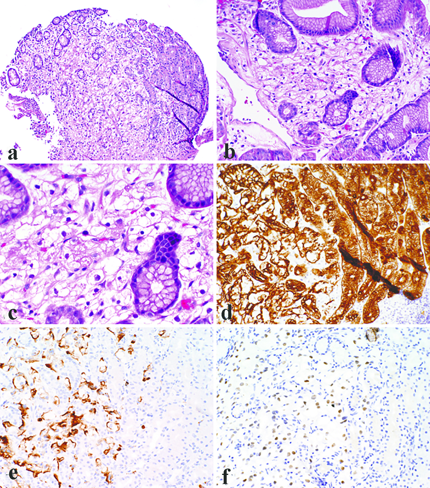
Figure 2. Representative photomicrographs of metastatic clear cell renal cell carcinoma as gastric mucosal lesions. The lesions demonstrated a bland clear cell proliferation within the lamina propria (a). At the interface between the carcinoma and benign gastric mucosa, the clear cells infiltrated among gastric glands without gland destruction (b). The individual tumor cells showed cytoplasmic vacuoles, small nuclei, nuclear membrane irregularity, and occasional small pinpoint nucleoli (c). Immunohistochemistry demonstrated immunoreactivity for pancytokeratin (d), RCC (e), and PAX8 (f). (a, × 100; b, d, e, f, × 200; c, × 400).
Figure 3.
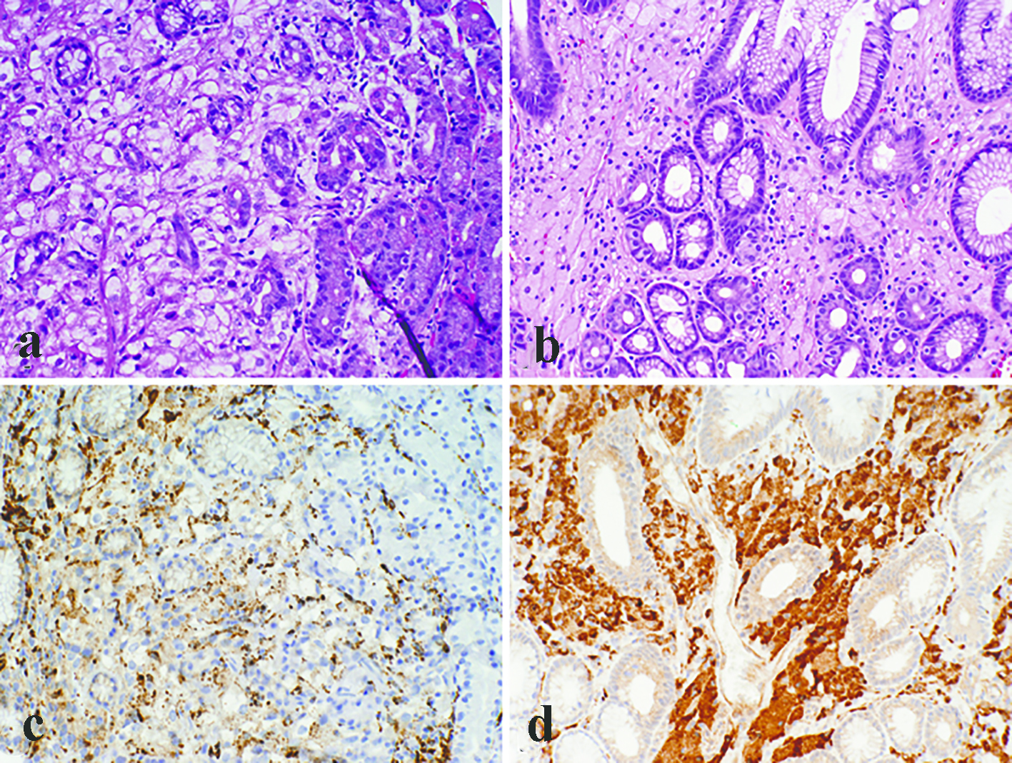
Figure 3. Metastatic renal cell carcinoma with many foamy histiocytes intermingled with the tumor cells (a). However, at least focal nuclear atypia was appreciated in the tumor cells, and immunohistochemistry for CD68 showed patchy staining (c). In contrast, gastric xanthomas were completely devoid of atypia and the cytoplasm was foamy rather than clear (b). Immunoreactivity for CD68 was strong and diffuse in xanthomas (d) (a, b, c, d, × 200).

Table

Table 1. Clinicopathologic Characteristics of Metastatic Renal Cell Carcinoma as Subcentimeter Polypoid Gastric Mucosal Lesions
 
Year*Age/sexSymptomsLocationSize (mm)Endoscopic characteristicsInterval#Primary stageOther sites of metastasesReference
GERD: gastroesophageal reflux disease; GI: gastrointestinal; N/A: no previous history of renal cell carcinoma. *Year of gastric metastasis for the current cases, year of publication for the referenced cases. #Years from the primary renal cell carcinoma diagnosis to the detection of gastric metastasis.
1201784/MDysphagia, GERDBody6Sessile polyp5pT1NxMxNoneCurrent
2201558/MOccasional GERDFundus4Sessile polyp6pT2N0MxAdrenal gland, pancreas, liver, lung (2012); brain (2015)Current
3201174/MUpper GI bleedingAntrum10Sessile polyp with ulcerated surfaceN/AUnknownNoneCurrent
4200872/FGERD, anemiaBody8Sessile polyp with ulcerated surfaceN/ApT2N0M1Liver, pancreas (2009)Current
5199866/MMelena, anemiaBody10Friable mass10pT1aN0MxLung, pancreas, mesentery (1998); brain (2000)Current
6201259/FGERD, back painFundus5SessileN/AUnknownBrain (unknown)[6]
7201260/FAnemiaBody6, 6Non-ulcerated, benign appearing polyps0.41pT1bNXLung, bone, and right nephrectomy bed (2011)[7]
8201279/MAbdominal painBody6Erosive lesionsynchronouspT1bN0M1None[8]
92007UnknownSevere anemia with melenaBody10Polypoid10UnknownNone[9]