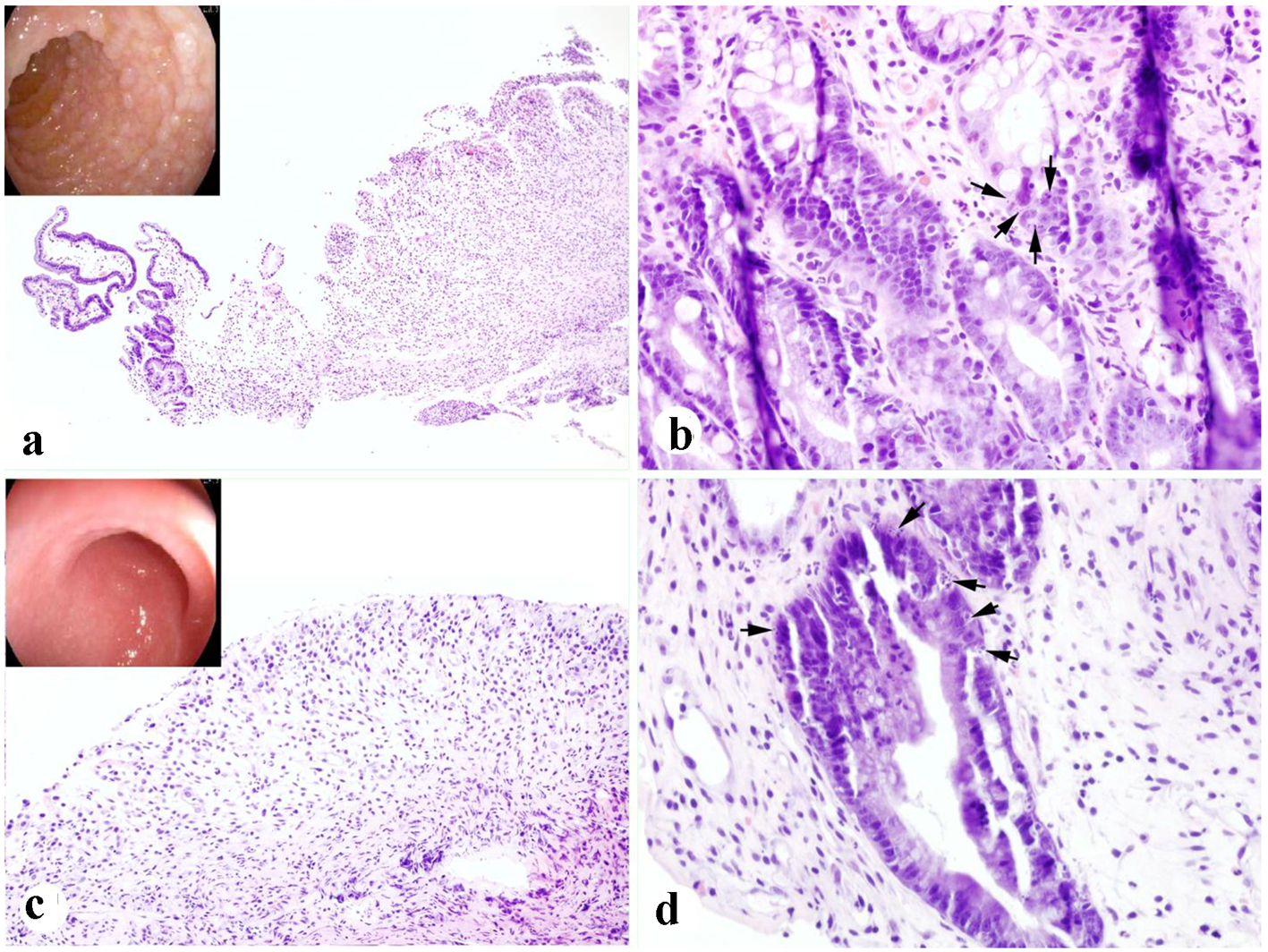
Figure 1. Severe duodenal and colonic auto-GVHD present in case 1. (a, b) Duodenal biopsy showing focal mucosal sloughing, crypt injury and dropout, and epithelial apoptotic bodies (arrows) (inset in a: endoscopic appearance). (c, d) Colonic biopsy showing extensive mucosal sloughing, crypt injury and dropout, and epithelial apoptotic bodies (arrows) (inset in c: endoscopic appearance) (H&E stain. a, c: × 100; b, d: × 400).
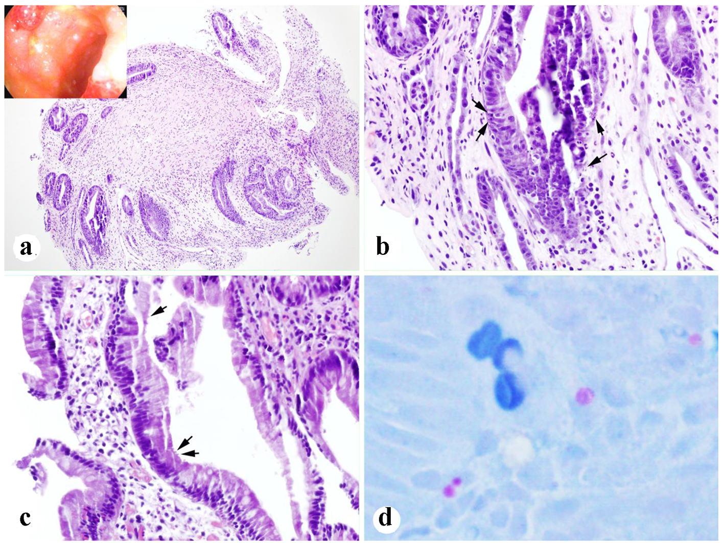
Figure 2. Biopsies after treatment for 13 days. (a, b) Colonic biopsy showing crypt regeneration and epithelial apoptotic bodies (arrows) (inset in a: endoscopic appearance). (c, d) Duodenal biopsy showing rare organisms of cryptosporidium (arrows) and highlighted by AFB (acid-fast) stain (d) (a: × 100; b, c: × 400; d: original, × 600).
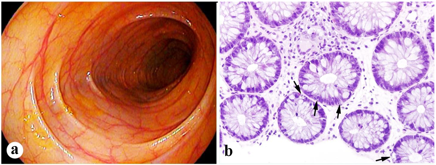
Figure 3. Mild colonic GVHD present in case 2. (a) Unremarkable endoscopic appearance of the colon. (b) Frequent apoptotic bodies present in the crypts of the colonic biopsy (arrows, H&E stain, × 400).