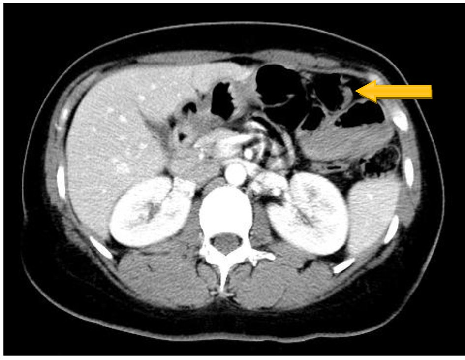
Figure 1. CT scan of abdomen shows dilated loop of small intestine in the left upper quadrant.
| Gastroenterology Research, ISSN 1918-2805 print, 1918-2813 online, Open Access |
| Article copyright, the authors; Journal compilation copyright, Gastroenterol Res and Elmer Press Inc |
| Journal website http://www.gastrores.org |
Case Report
Volume 10, Number 6, December 2017, pages 366-368
Primary Jejunal Adenocarcinoma Presenting as Bilateral Ovarian Metastasis
Figures