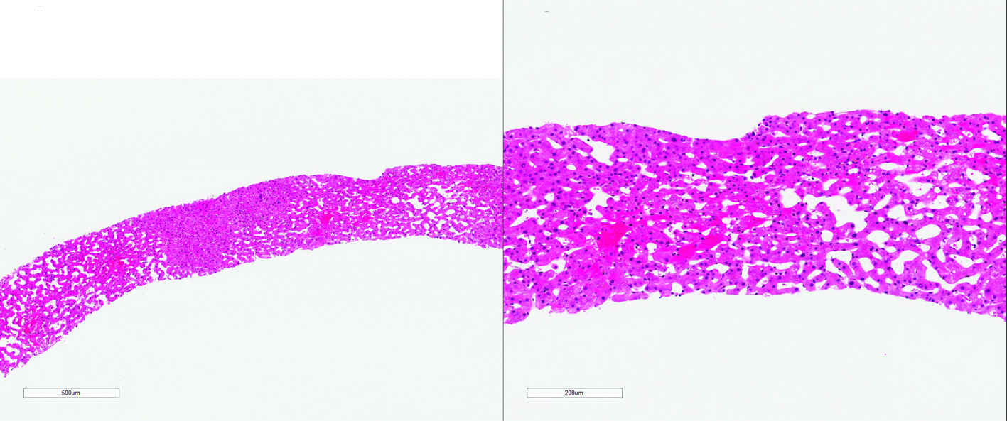
Figure 1. Morphologic features seen in congestive hepatopathy, including marked sinusoidal dilatation, centrilobular hepatocellular atrophy and hemorrhage (H&E).
| Gastroenterology Research, ISSN 1918-2805 print, 1918-2813 online, Open Access |
| Article copyright, the authors; Journal compilation copyright, Gastroenterol Res and Elmer Press Inc |
| Journal website http://www.gastrores.org |
Original Article
Volume 10, Number 3, June 2017, pages 182-189
Histology and Glutamine Synthetase Immunoreactivity in Liver Biopsies From Patients With Congestive Heart Failure
Figures
Tables
| Fibrosis stage | Definition | Score/code |
|---|---|---|
| No fibrosis | 0 | |
| Central zone fibrosis | 1 | |
| Central zone and mild portal fibrosis, with accentuation at central zone | 2A | |
| Central zone and mild portal fibrosis, with accentuation at portal zone | 2B | |
| Bridging fibrosis | 3 | |
| Cirrhosis | 4 | |
| Central zone fibrosis | ||
| No fibrosis | Absent | |
| Presence of fibrosis | Present | |
| Sinusoidal dilatation | ||
| Normal width of sinusoids | Absent | |
| Widening of sinusoids | Present | |
| Centrilobular hemorrhage | ||
| Red blood cells confined in sinusoids | Absent | |
| Red blood cells beyond sinusoids | Present | |
| Hepatocyte atrophy | ||
| Normally sized hepatocytes | Absent | |
| Hepatocyte smaller than normal hepatocytes | Present | |
| Steatosis grade | ||
| No steatosis | Absent | |
| (< 5% of biopsy volume) | Minimal | |
| (5-33% of biopsy volume) | Mild | |
| (34-66% biopsy volume) | Moderate | |
| (≥ 67% of biopsy volume) | Severe | |
| Cholestasis | ||
| No cholestasis | Absent | |
| Cholestasis in hepatocytes, canaliculi, or cholangioles | Present | |
| Hepatocyte ballooning | ||
| No ballooning in any hepatocytes | Absent | |
| Enlarged round hepatocytes with rarefication of cytoplasm | Present | |
| Mallory hyalines | ||
| No | Absent | |
| Any intrahepatocytic ropy eosinophilic filaments | Present | |
| Ductular proliferation | ||
| Normal bile duct profile | Absent | |
| Duct-like structure with or without lumen at the periphery of limiting plate | Present | |
| Polymorphonuclear leukocyte inflammation | ||
| No neutrophilic inflammation | Absent | |
| Rare neutrophils in the portal tracts and the lobules | Present | |
| Mononuclear inflammation | ||
| No mononuclear inflammation | Absent | |
| Mononuclear inflammatory cells in the sinusoids, portal tracts, or in the lobules | Present |
| N (%) | |
|---|---|
| Stage of fibrosis | |
| No fibrosis | 5 (14) |
| Centrilobular fibrosis only (1) | 4 (11) |
| Centrilobular (prominent) and portal fibrosis (2A) | 6 (17) |
| Centrilobular and portal (prominent) fibrosis (2B) | 5 (14) |
| Bridging fibrosis (3) | 6 (17) |
| Cirrhosis (4) | 4 (11) |
| Not cirrhosis, but with fibrosis of indeterminate degree | 6 (17) |
| Centrilobular fibrosis | |
| Absent | 6 (17) |
| Present | 25 (69) |
| Indeterminate | 5 (14) |
| Sinusoidal dilatation | |
| Absent | 8 (22) |
| Present | 26 (72) |
| Indeterminate | 2 (6) |
| Hepatocyte atrophy | |
| Absent | 12 (34) |
| Present | 21 (58) |
| Indeterminate | 3 (8) |
| Centrilobular hemorrhage | |
| Absent | 15 (42) |
| Present | 17 (47) |
| Indeterminate | 4 (11) |
| Steatosis | |
| < 5% | 34 (94) |
| 5-33% | 1 (3) |
| 33-66% | 0 (0) |
| > 66% | 1 (3) |
| Ballooning | |
| Absent | 35 (97) |
| Present | 1 (3) |
| Mallory hyalines | |
| Absent | 36 (100) |
| Present | 0 (0) |
| Cholestasis | |
| Absent | 35 (97) |
| Present | 1(3) |
| Lobular polymorphonuclear leukocyte infiltration | |
| Absent | 36 (100) |
| Present | 0 (0) |
| Lobular mononuclear inflammation | |
| Absent | 21 (58) |
| Present | 13 (36) |
| Indeterminate | 2 (6) |
| Portal polymorphonuclear leukocyte infiltration | |
| Absent | 35 (97) |
| Present | 1 (3) |
| Portal mononuclear inflammation | |
| Absent | 10 (28) |
| Present | 19 (53) |
| Indeterminate | 7 (19) |
| Ductular proliferation | |
| Absent | 7 (20) |
| Present | 13 (36) |
| Indeterminate | 16 (44) |
| Histologic feature | K value (95% CI) | Strength of agreement |
|---|---|---|
| Sinusoidal dilation (absent/present) | 0.66 (0.41 - 0.86) | Substantial |
| Centrilobular atrophy (absent/present) | 0.51 (0.35 - 0.70) | Moderate |
| Centrilobular hemorrhage (absent/present) | 0.35 (0.19 - 0.53) | Fair |
| Centrilobular fibrosis (absent/present) | 0.43 (0.21 - 0.68) | Moderate |
| Fibrosis stage (congestive hepatic fibrosis score) | 0.35 (0.24 - 0.49) | Fair |
| Simplified fibrosis stage (CHFS 0-2B/3-4) | 0.45 (0.31 - 0.61) | Moderate |
| Histologic feature | Correlation coefficient | Significance (P value) |
|---|---|---|
| Sinusoidal dilation (absent/present) | 0.209 | 0.317 |
| Centrilobular atrophy (absent/present) | 0.092 | 0.66 |
| Centrilobular hemorrhage (absent/present) | 0.192 | 0.356 |
| Centrilobular fibrosis (absent/present) | -0.126 | 0.546 |
| Fibrosis stage (congestive hepatic fibrosis score) | 0.107 | 0.571 |