Gastroenterology Research, ISSN 1918-2805 print, 1918-2813 online, Open Access
Article copyright, the authors; Journal compilation copyright, Gastroenterol Res and Elmer Press Inc
Journal website http://www.gastrores.org

Case Report

Volume 10, Number 4, August 2017, pages 255-258


Intraductal Ultrasonography as a Local Assessment Before Magnetic Compression Anastomosis for Obstructed Choledochojejunostomy

Figures

Figure 1.
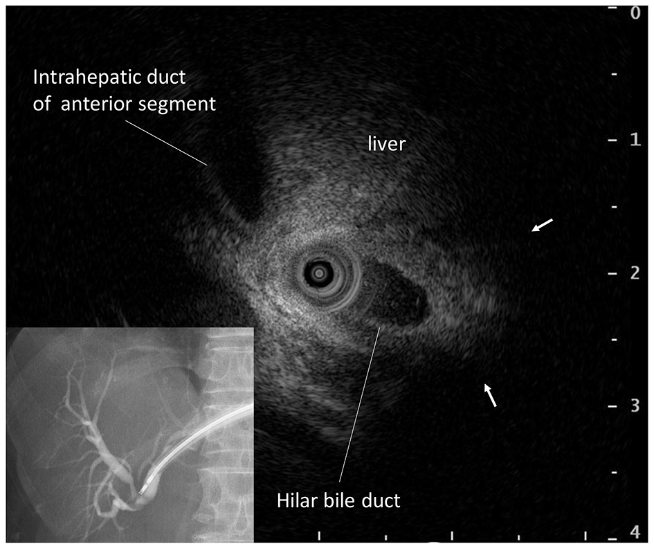
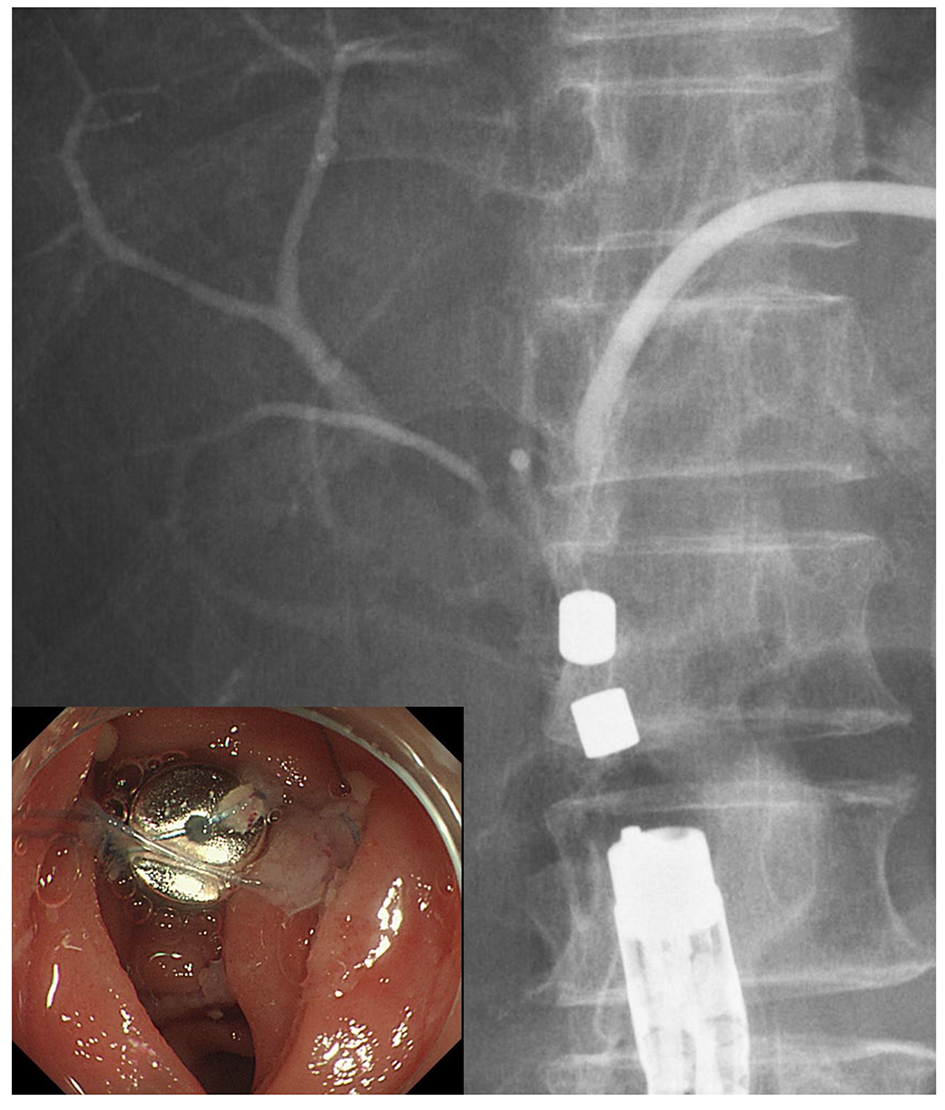
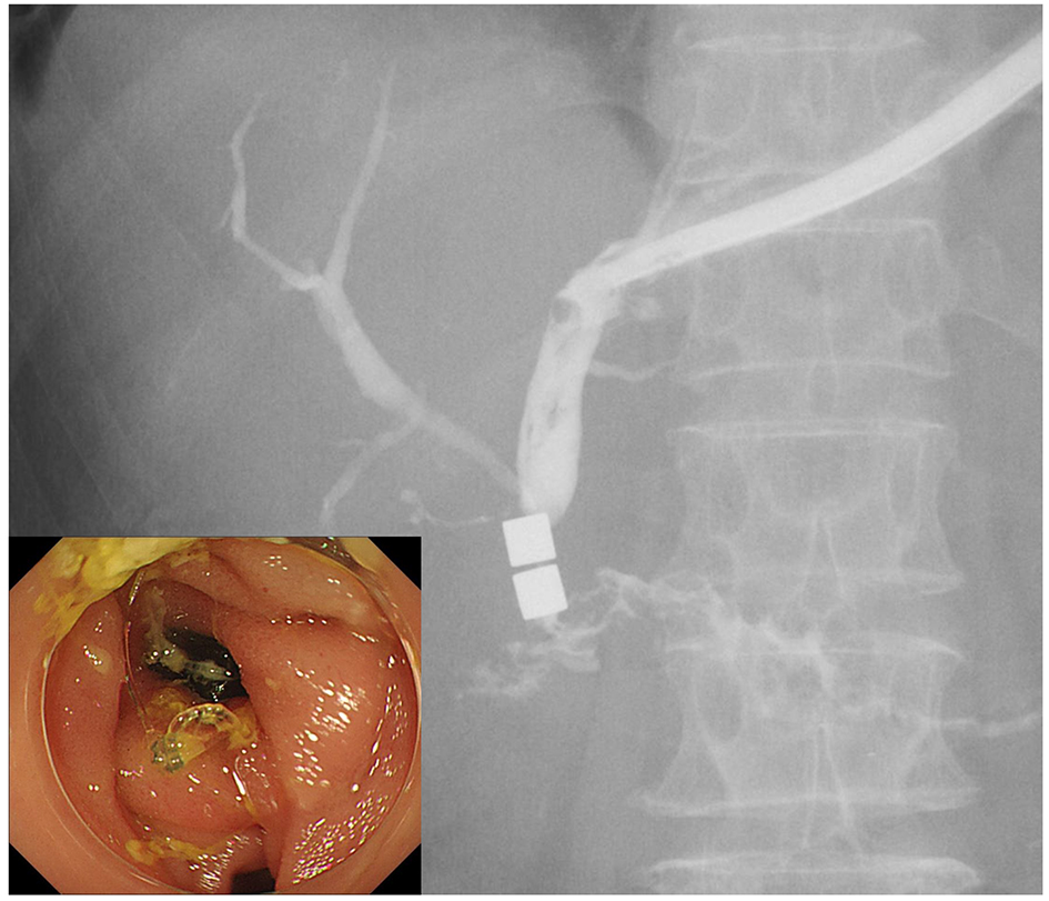
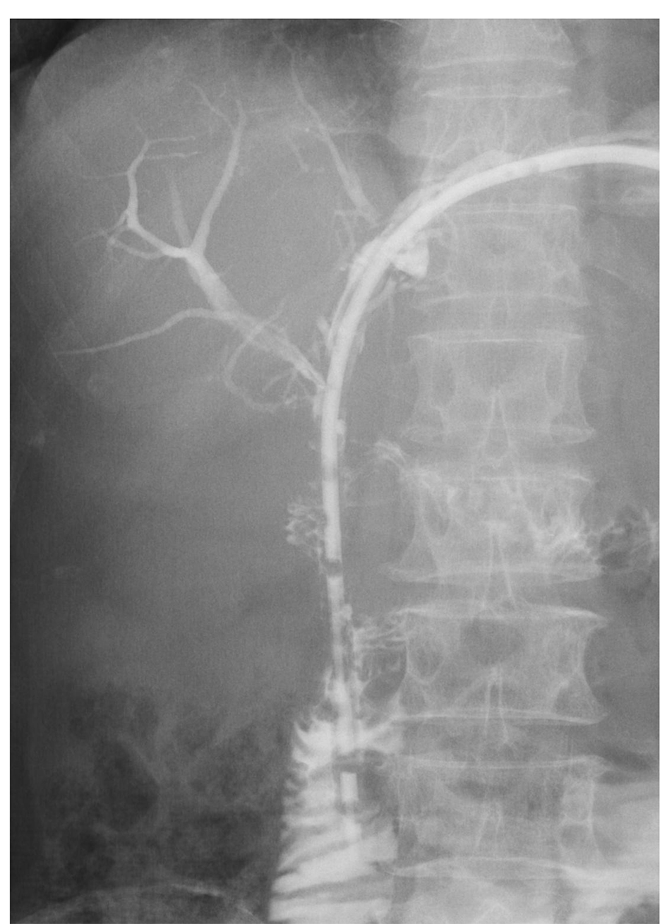
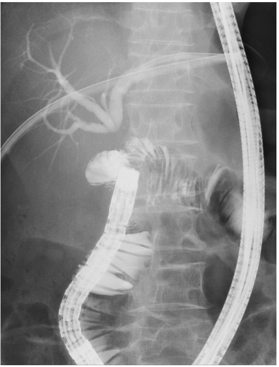
Figure 1. Percutaneous transhepatic cholangiography showed complete obstruction of choledochojejunostomy. The obstructive distance was 7 mm, as measured by cholangiography and fluoroscopy.
Figure 2.
Figure 2. Intraductal ultrasonography through the percutaneous transhepatic cholangiographic drainage tube revealed a fibrous heterogeneous hyperechoic appearance without fluid collection, vessels or foreign bodies between the common bile duct and jejunum (arrow).
Figure 3.
Figure 3. The two magnets were attracted towards each other transmurally and compressed the obstruction site from both sides.
Figure 4.
Figure 4. After reanastomosis was confirmed, the magnets were retrieved endoscopically.
Figure 5.
Figure 5. A 16-Fr indwelling drainage tube was placed in the juodenum through percutaneous transhepatic cholangiographic drainage tube.