Gastroenterology Research, ISSN 1918-2805 print, 1918-2813 online, Open Access
Article copyright, the authors; Journal compilation copyright, Gastroenterol Res and Elmer Press Inc
Journal website http://www.gastrores.org

Case Report

Volume 10, Number 3, June 2017, pages 208-211


Two Cases of Pneumatosis Cystoides Intestinalis With Intraperitoneal Free Air

Figures

Figure 1.
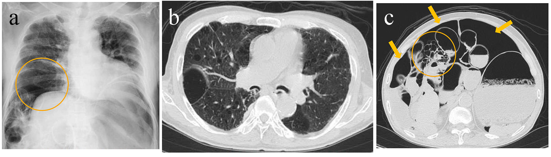
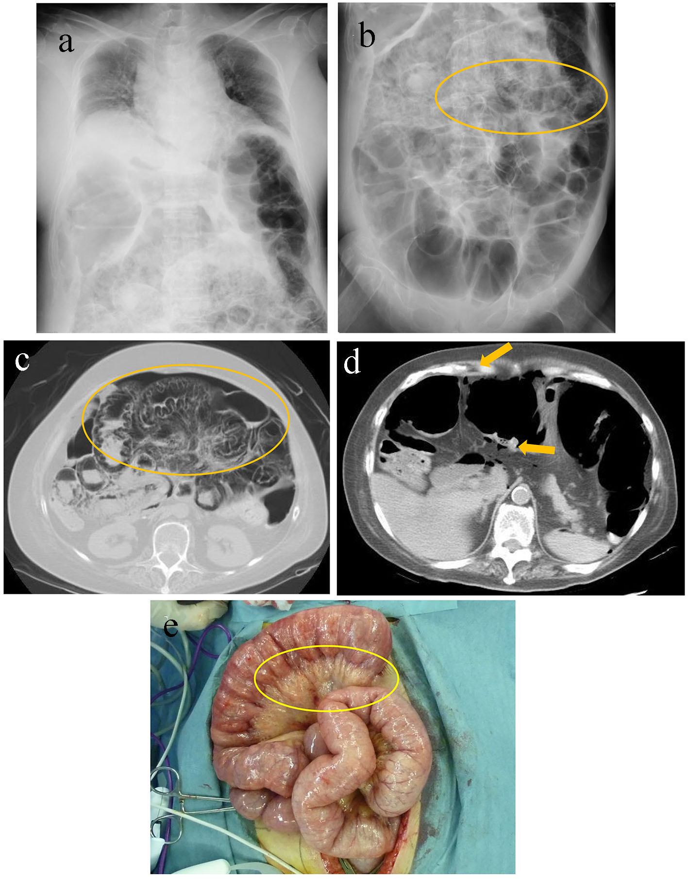
Figure 1. A chest radiography revealed elevation of the diaphragm due to dilated and gaseous intestines (a). A radiograph of the abdomen revealed diffusely dilated intestines and multiple, small radiolucent linear bubbles lining the intestines (circle) (b). Abdominal computed tomography revealed dilated and gaseous small intestines, retention of gas in the intestinal wall (circle) (c), and intraperitoneal free air (arrows) (d). Operative findings revealed small air bubbles in the intestinal wall and mesentery (e).
Figure 2.
Figure 2. A chest radiography revealed decompression of the right lung with an elevated diaphragm and the retention of massive free air under the diaphragm (circle) (a). Non-contrast computed tomography revealed bilateral multiple emphysematous bullae in the lungs (b), distended small intestines, and intraperitoneal free air (arrows) along with multiple, small bubbly cysts beneath the intestinal serosa (circle) (c).