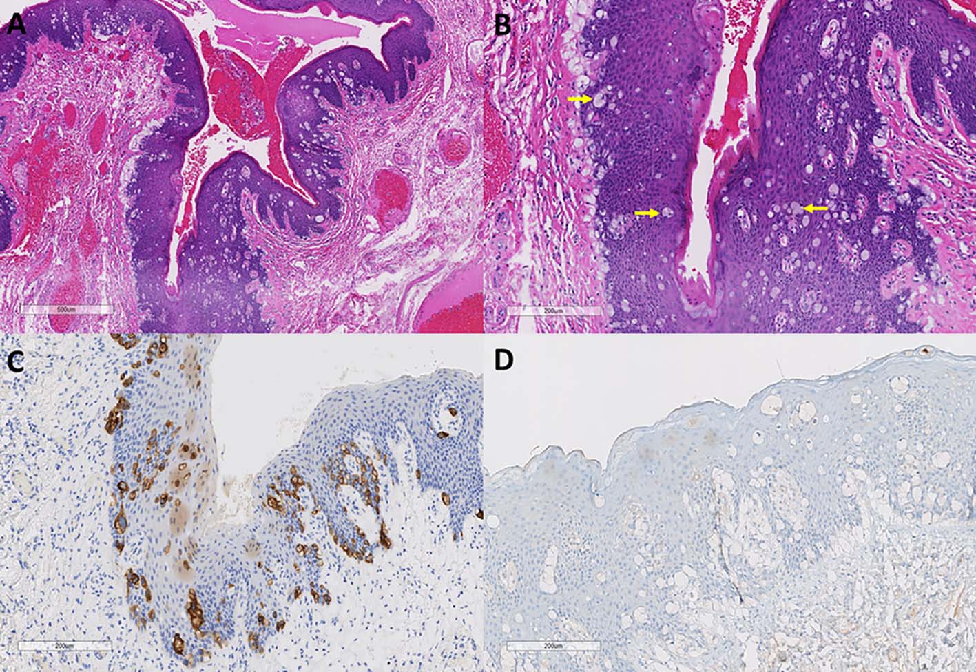
Figure 1. Paget’s disease of anal canal. (A, B) Anal mucosa from first resection specimen showing infiltration of large atypical vacuolated cells (Paget’s cells, arrows) with cytoplasmic mucin (hematoxylin and eosin stain, original magnification × 40 (A); × 100 (B)). (C, D) These cells are CK20 positive and GCDFP15 negative (immunoperoxidase stain, CK20, original magnification × 100 (C); GCDFP15, original magnification × 100 (D)).
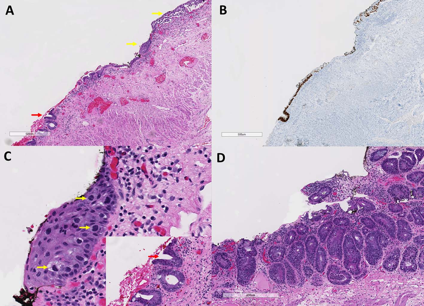
Figure 2. Residual Paget’s disease and rectal adenoma. (A) Anal mucosa from second resection specimen showing residual Paget’s disease (yellow arrows) adjacent to flat adenoma (red arrow) (hematoxylin and eosin stain, original magnification, × 40). (B) The Paget’s cells and adenomatous cells are CDX2 positive (immunoperoxidase stain, CDX2, original magnification, × 40). (C) Higher magnification view of residual Paget’s disease (yellow arrows) and adenomatous crypts (inset with red arrow) (hematoxylin and eosin stain, original magnification, × 200). (D) Adenoma in the smaller tissue fragment (hematoxylin and eosin, original magnification, × 62).