Gastroenterology Research, ISSN 1918-2805 print, 1918-2813 online, Open Access
Article copyright, the authors; Journal compilation copyright, Gastroenterol Res and Elmer Press Inc
Journal website http://www.gastrores.org

Case Report

Volume 7, Number 5-6, December 2014, pages 146-148


Hypervascular Lesion in a Cirrhotic Liver: A Case Report

Figures

Figure 1.
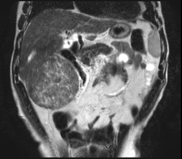
Figure 1. Abdominal contrasted MRI, T2-weighted, showing the tumor. It presented close proximity to the right kidney and the hepatic flexure of the colon.
Figure 2.
Figure 2. Intraoperative picture of the tumor.
Figure 3.
Figure 3. Tumor sectioned in half.
Figure 4.
Figure 4. Image of the tumor sectioned in half, with detail to one of its nodules. It is possible to observe a nodule that microscopic analyses could not conclude whether it was completely extirpated from the patient.
Figure 5.
Figure 5. Microscopic view of the part of the tumor where the nodule was sectioned. It is possible to observe that cancerous cells were present in the nodule, and there is no margin of normal cells, which leads us to believe that there are still cancerous cells in the organ.