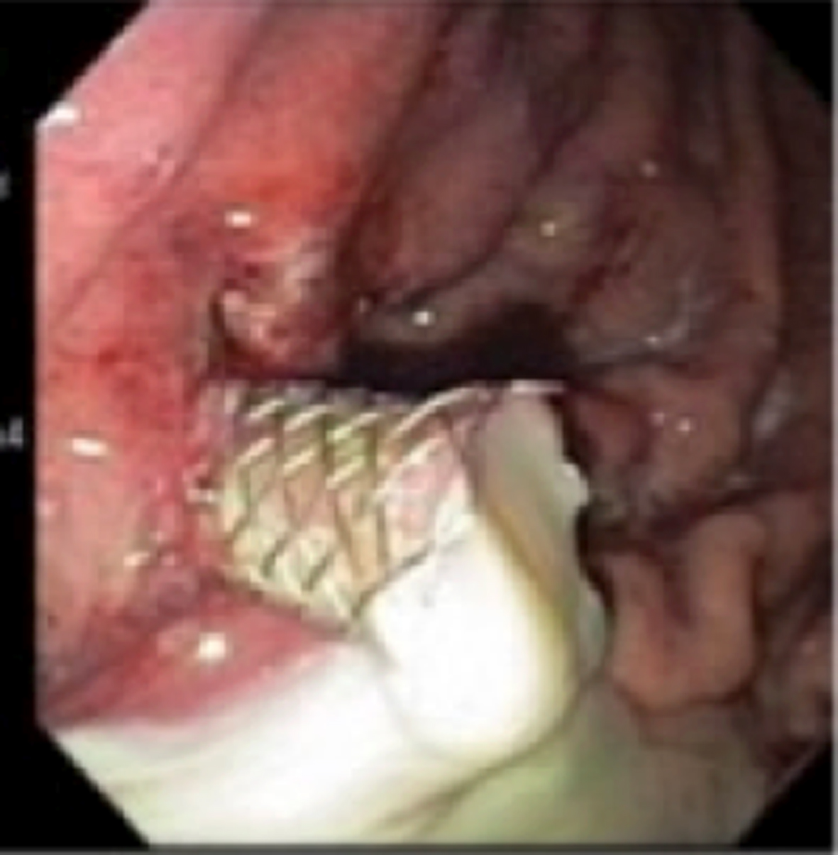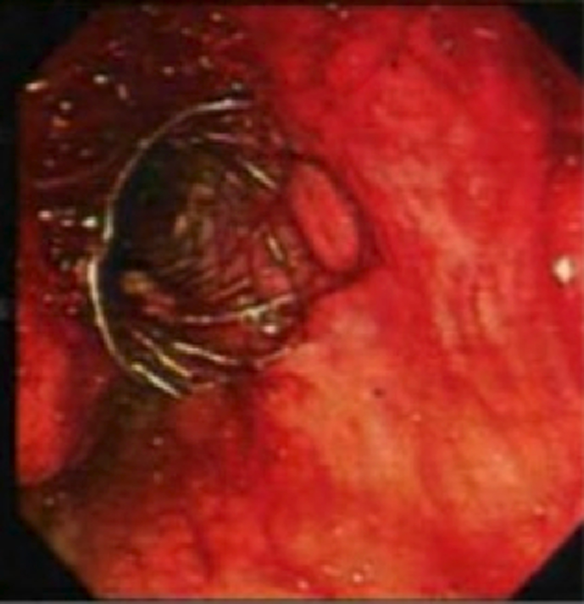
Figure 1. SEMS placed via the transgastric approach.
| Gastroenterology Research, ISSN 1918-2805 print, 1918-2813 online, Open Access |
| Article copyright, the authors; Journal compilation copyright, Gastroenterol Res and Elmer Press Inc |
| Journal website http://www.gastrores.org |
Case Report
Volume 7, Number 3-4, August 2014, pages 105-110
Endoscopic Ultrasound-Guided Self-Expandable Metal Stent Placement for the Treatment of Infected Pancreatic Pseudocysts
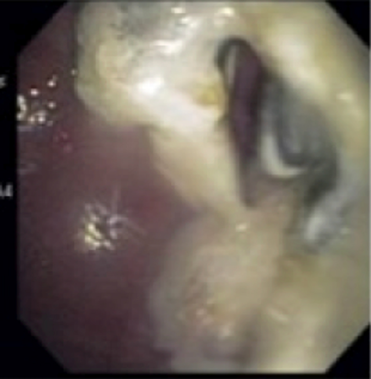
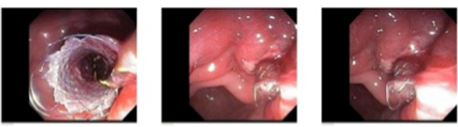
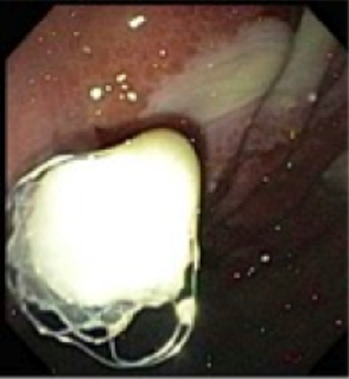
Figures