Gastroenterology Research, ISSN 1918-2805 print, 1918-2813 online, Open Access
Article copyright, the authors; Journal compilation copyright, Gastroenterol Res and Elmer Press Inc
Journal website http://www.gastrores.org

Case Report

Volume 7, Number 2, April 2014, pages 69-72


Acute Hepatitis E in the US Today Occurs in Diverse Patient Populations: Case Reports

Figures

Figure 1.
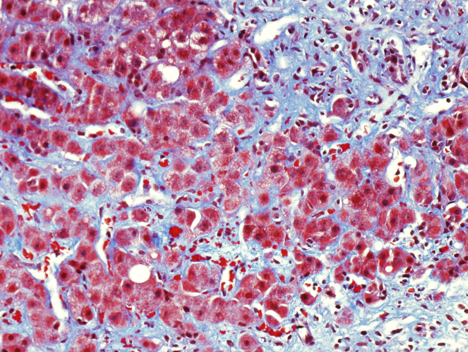
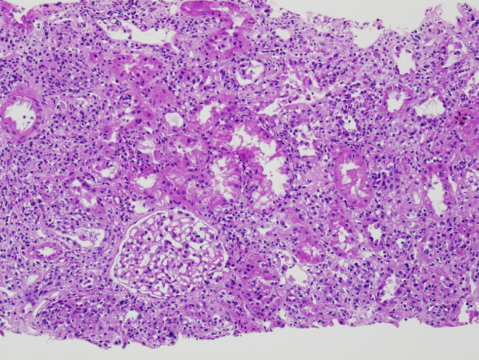
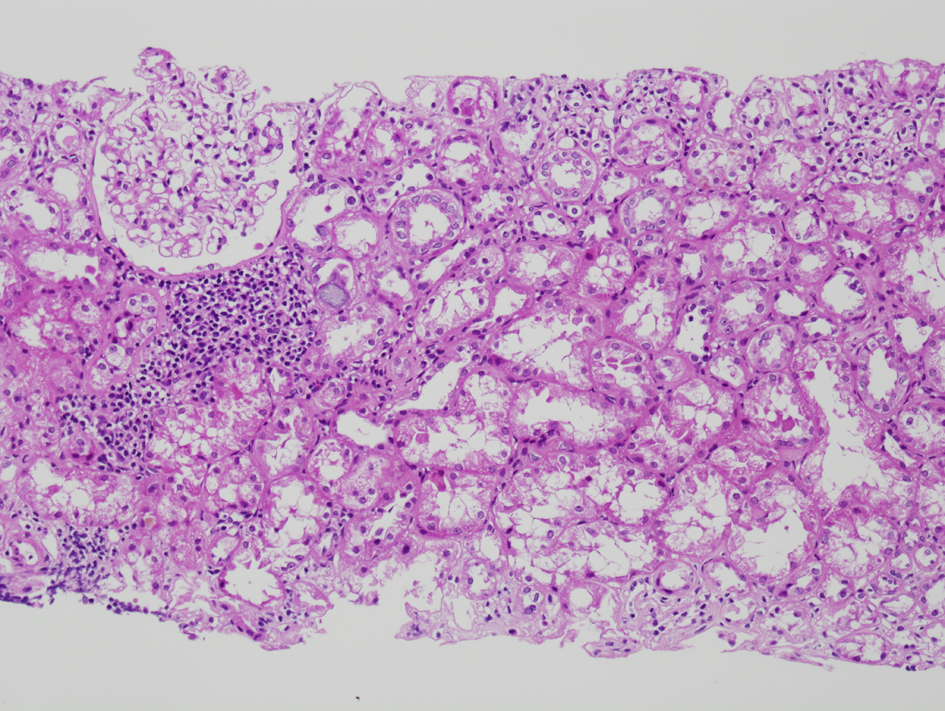
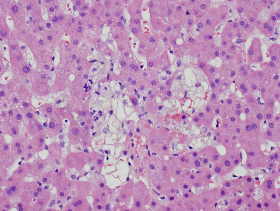
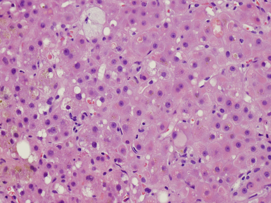
Figure 1. Initial liver biopsy showing lobular hepatitis, focal mild canalicular and intracellular cholestasis, focal intracellular fibrinogen, and mild steatosis (H and E, × 200).
Figure 2.
Figure 2. Follow-up liver biopsy, trichrome stain showing marked portal and perisinusoidal fibrosis.
Figure 3.
Figure 3. Initial renal biopsy showing a small discrete focus of interstitial lymphohistiocytic inflammation (H and E, × 100).
Figure 4.
Figure 4. Repeat renal biopsy showing marked diffuse interstitial infiltration by lymphocytes (H and E, × 100). On immunochemical stain (not shown here), the lymphocytes were predominantly CD3 positive T cells with a predominance of CD8 positive T cells.
Figure 5.
Figure 5. Liver biopsy showing focal hepatocyte dropout (H and E, × 200)