Figures
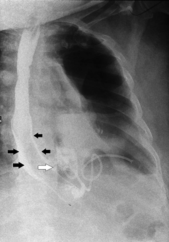
Figure 1. Contrast study of esophagus: no contrast is seen going across the lower end of stent. Contrast is seen seeping along the sides of the SEMS (black arrow) and then leaking into pleural cavity (white arrow). A pigtail is seen inside the left pleural cavity.
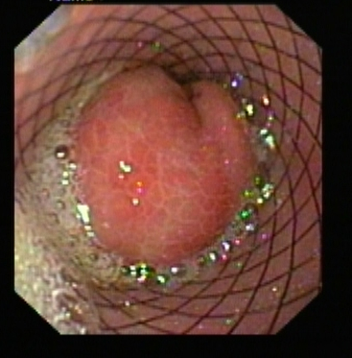
Figure 2. Lower end of SEMS blocked by prolapsed gastric mucosa.
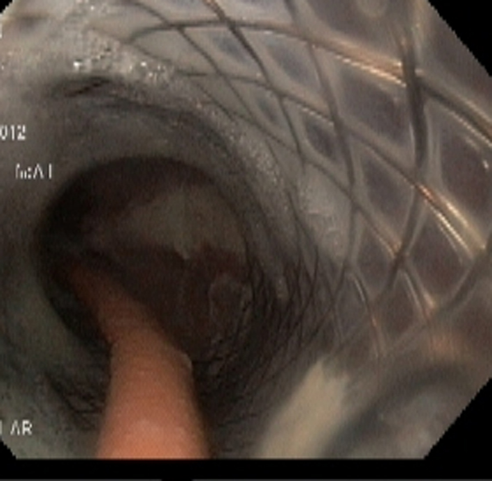
Figure 3. Endoscopy done 3 weeks after placement of NJT. The NJT is seen going through the SEMS and lower end of SEMS is seen opened up.
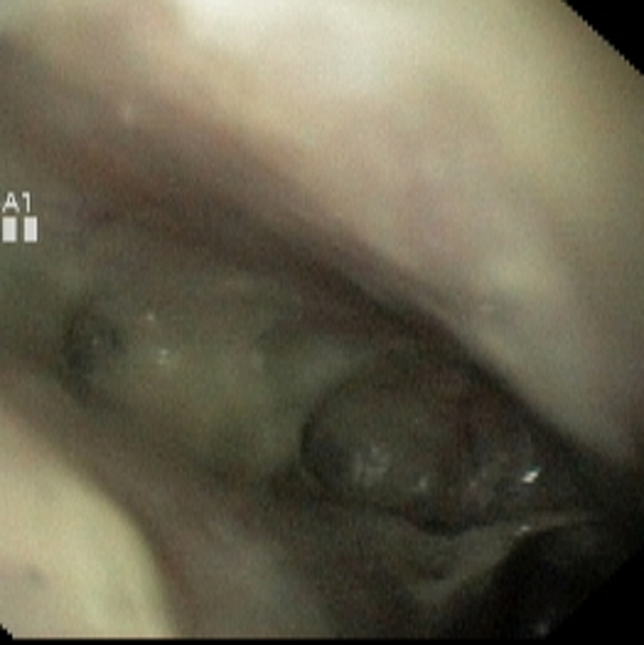
Figure 4. Large perforation at lower end of esophagus.
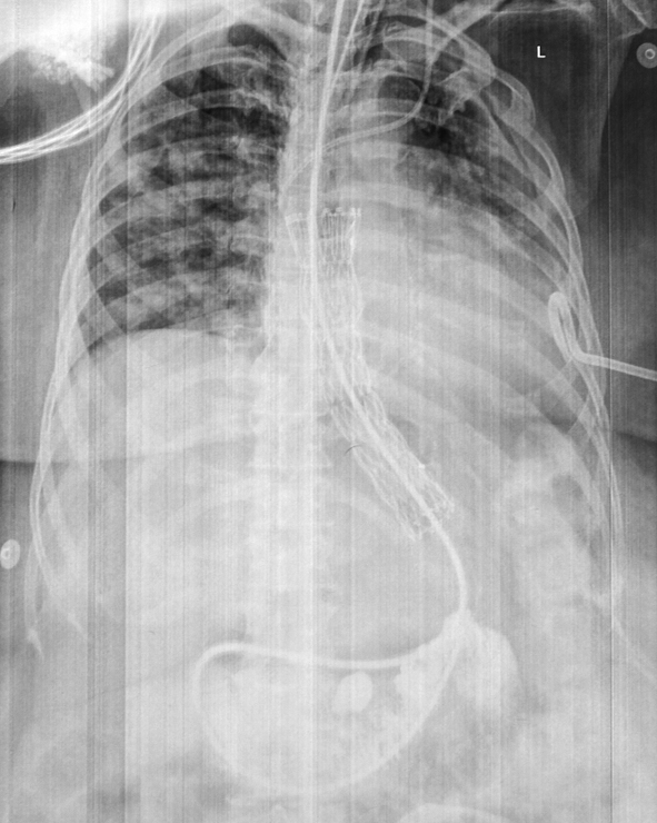
Figure 5. NJT is seen passing through SEMS into the jejunum. Pigtail in left pleural cavity and central line catheter are also noted.
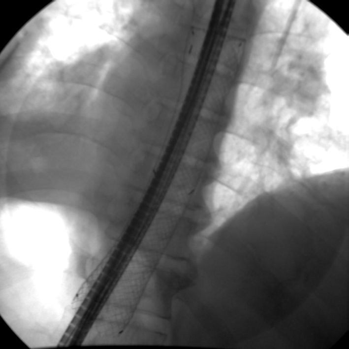
Figure 6. NJT placed through the SEMS.
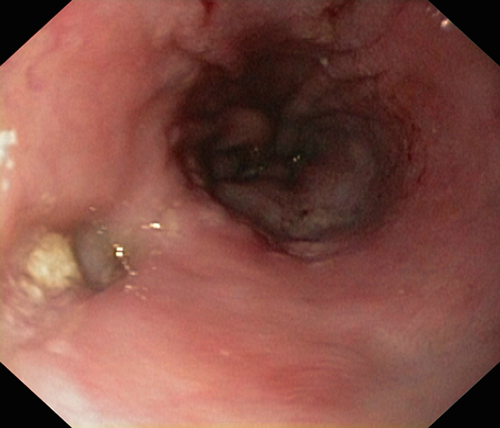
Figure 7. Endoscopy after stent removal: small depression is noted at the site of perforation.
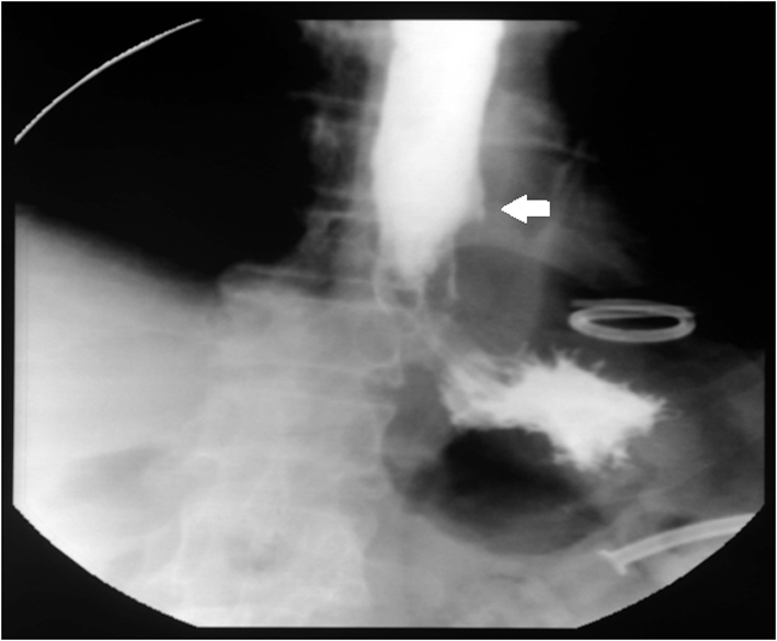
Figure 8. Contrast study after stent removal: no leakage of contrast is seen and a small outpouching is seen at site of perforation (arrow).