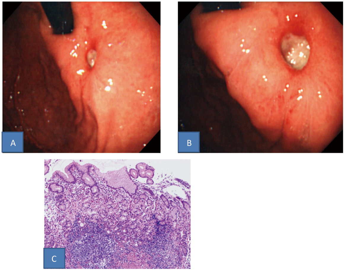
Figure 1. A, B: Upper gastrointestinal endoscopic findings (A: Intermediate view. B: Near view). A small, uedermined ulcer was found in the gastric cardia. C: Histopathological findings in a biopsy specimen (HE stain). Specific findings were not found: only hyperplastic changes of the cryptic epithelium and inflammatory cell infiltration in the storoma.
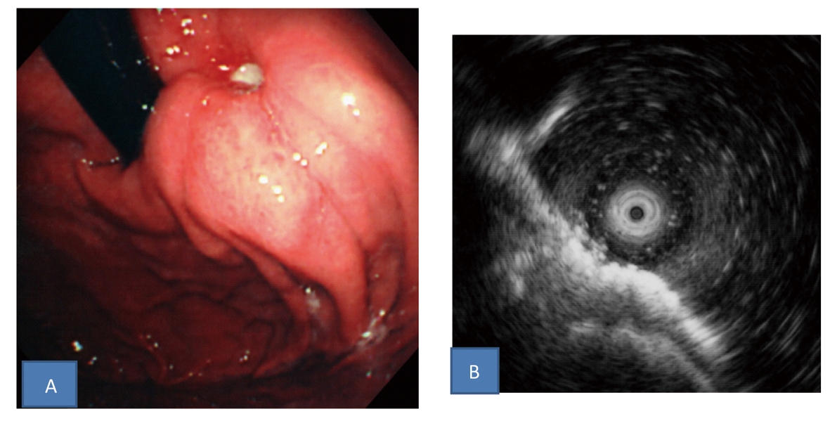
Figure 2. A: Upper gastrointestinal endoscopic findings after Hp eradication therapy followed by 8 weeks of PPI administration. The ulcer slightly shrunk without scarring and the surrounding mucosa was markedly elevated like a submucosal tumor. B: Endoscopic ultrasonography findings. Thickening of the submucosal and muscular layers around the ulcer was found.
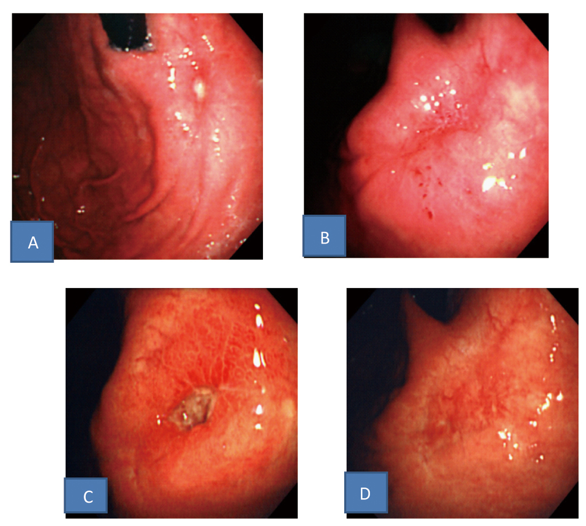
Figure 3. Upper gastrointestinal endoscopic findings. A: The ulcer shrunk and the elevation around the ulcer disappeared after continuous PPI administration. B: The ulcer scarred after more continuous PPI administration. C: The ulcer recurred after PPI dose reduction in spite of successful Hp eradication. D: The ulcer scarred again after increase of PPI dosage.