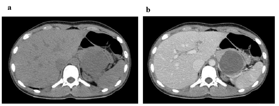
Figure 1. CT images at 4 h after abdominal injury. (a) Plain CT reveals a cystic mass measuring 55 mm in the pancreatic body and tail. (b) Contrast-enhanced CT indicates a heterogeneous internal density and smooth rim enhancement of the mass.
| Gastroenterology Research, ISSN 1918-2805 print, 1918-2813 online, Open Access |
| Article copyright, the authors; Journal compilation copyright, Gastroenterol Res and Elmer Press Inc |
| Journal website http://www.gastrores.org |
Case Report
Volume 6, Number 2, April 2013, pages 67-70
A Pancreatic Solid Pseudo-Papillary Tumor Detected After Abdominal Injury
Figures
Table
| Item | Value | Unit |
|---|---|---|
| WBC | 11.2 | 103/µL |
| Neutrophil | 85.8 | % |
| Lymphocyte | 11.1 | % |
| RBC | 484 | 104/µL |
| Hb | 13.4 | g/dL |
| Plt | 23.5 | 104/µL |
| Protein | 8.0 | g/dL |
| Albumin | 4.8 | g/dL |
| UN | 12 | mg/dL |
| Cr | 0.6 | mg/dL |
| Na+ | 141 | mmol/L |
| K+ | 4.4 | mmol/L |
| Cl− | 104 | mmol/L |
| Ca2+ | 9.6 | mg/dL |
| Total bilirubin | 0.3 | mg/dL |
| AST | 17 | IU/L |
| ALT | 17 | IU/L |
| ALP | 411 | IU/L |
| γ-GTP | 14 | IU/L |
| CK | 315 | IU/L |
| Amylase | 287 | U/L |
| Glucose | 130 | mg/dL |
| CRP | 0.0 | mg/dL |
| PT | 11.1 | sec |
| APTT | 24.9 | sec |
| Fibrinogen | 292 | mg/dL |
| D-dimer | < 0.5 | mg/mL |
| CAE | 0.8 | ng/mL |
| CA19-9 | 11 | U/mL |
| AFP | 1.7 | ng/mL |
| NSE | 22 | ng/mL |