Gastroenterology Research, ISSN 1918-2805 print, 1918-2813 online, Open Access
Article copyright, the authors; Journal compilation copyright, Gastroenterol Res and Elmer Press Inc
Journal website http://www.gastrores.org

Case Report

Volume 6, Number 2, April 2013, pages 63-66


Bleeding Brunner’s Gland Hyperplasia of the Duodenum: A Case Report With a Review of the Japanese Literature

Figures

Figure 1.
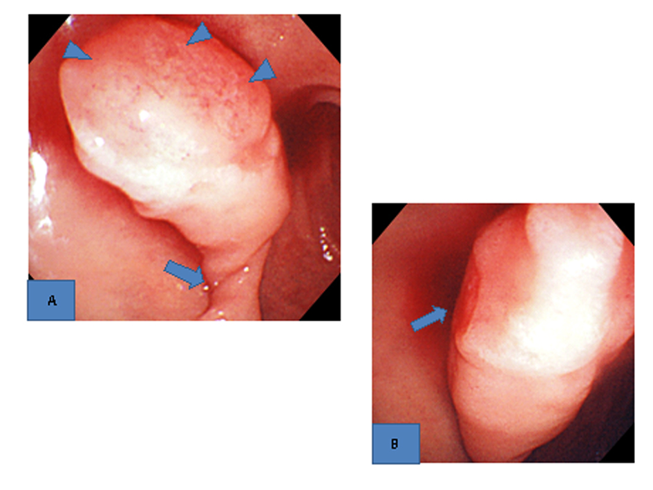
Figure 1. Upper gastrointestinal endoscopic findings. A). A large pedunculated poly with extensive erosions on the top (arrowheads) and a twisted pedicle (arrow) was found in the duodenal bulb. B). Oozing hemorrhage from the erosions on the top was seen (arrow).
Figure 2.
Figure 2. Upper gastrointestinal endoscopic findings. A). The bleeding from the erosions on the top stopped. B). The twisting of the pedicle was reduced. C). Resected specimen. A thick blood vessel was found in the pedicle.
Figure 3.
Figure 3. Histopathological findings (H.E. stain). A). The surface of the polyp was covered with an eroded epithelium, and hyperplastic Brunner’s glands were densely packed in the submucosal layer (low-power view). B). Atypia was not found (high-power view).

Table

Table 1. Reported Cases of Bleeding Burnner’s Hyperplasia in Japan (1996 - 2010, Including Our Case)
 
Number of cases2
Age: 40 y.o. ≥ / 40 y.o. <7/2
Sex: male/female7/2
Form: pedunculated polyp/others9/0
Size: 2 cm ≥ / 2 cm <8/1
Location: bulb/descending part8/1
Antithrombotic drugs: (+)/(-)2/7