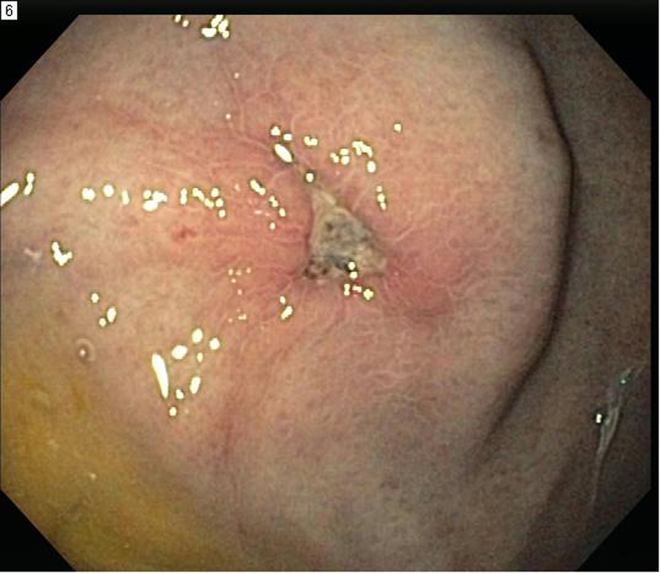
Figure 1. Endoscopic image showing large antral ulcer with central red pigmentation and surrounding erythema.
| Gastroenterology Research, ISSN 1918-2805 print, 1918-2813 online, Open Access |
| Article copyright, the authors; Journal compilation copyright, Gastroenterol Res and Elmer Press Inc |
| Journal website http://www.gastrores.org |
Case Report
Volume 6, Number 1, February 2013, pages 26-28
Gastroduodenal Cryptococcus in an AIDS Patient Presenting With Melena
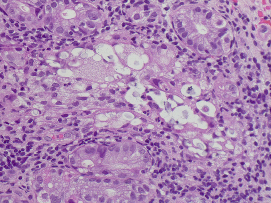
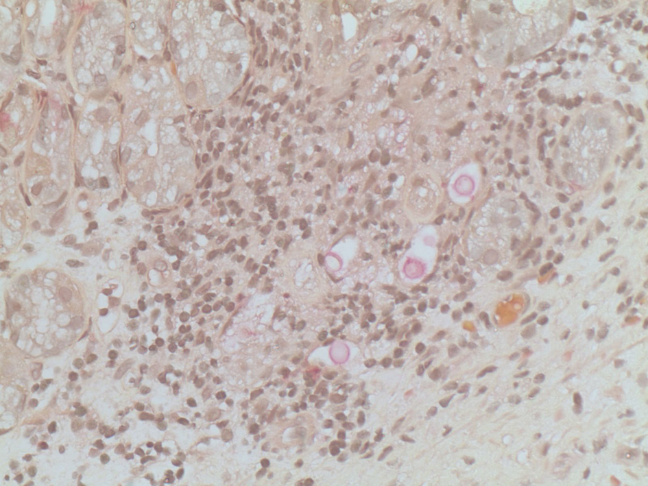
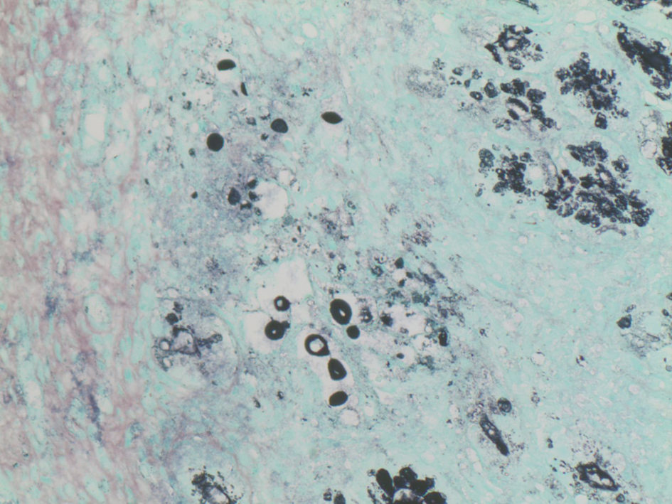
Figures