Gastroenterology Research, ISSN 1918-2805 print, 1918-2813 online, Open Access
Article copyright, the authors; Journal compilation copyright, Gastroenterol Res and Elmer Press Inc
Journal website http://www.gastrores.org

Case Report

Volume 5, Number 6, December 2012, pages 245-248


Gallbladder Papillary Neoplasia Associated With Intrahepatic Carcinoma and Pancreaticobiliary Malformation

Figures

Figure 1.
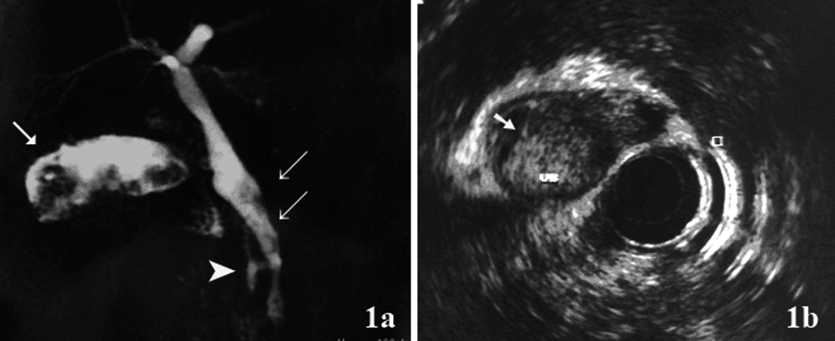
Figure 1. (a) Magnetic resonance cholangiopancreatography (MRCP) showed hyperdense structure within the gallbladder infundibulum, which were suggestive of tumor (arrow), bile duct dilatation and filling defect (double arrow) and pancreaticobiliary maljunction(arrow head); (b) Endoscopic ultrasound showed hyperechoic frond-like mass within the gallbladder (arrow).
Figure 2.
Figure 2. Cancer of the gallbladder and the bile duct. (a) Tumor within the gallbladder and the lumen was filled with mucin. (b, c) Papillary adenocarcinoma extending to the superficial portion of the muscle layer (H&E, × 25 and × 50). (d) The specimen of the liver showed carcinoma in situ in the segmental bile duct (H&E, × 100).