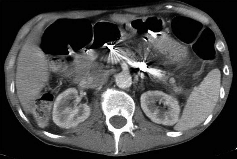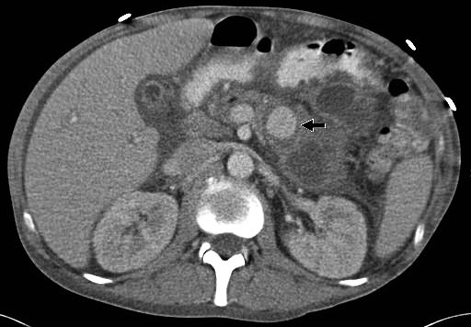
Figure 1. Pre-procedure CTA demonstrating diffuse pancreatitis with pseudocyst and a large pseudoaneurysm arising from a branch of the superior mesenteric artery.
| Gastroenterology Research, ISSN 1918-2805 print, 1918-2813 online, Open Access |
| Article copyright, the authors; Journal compilation copyright, Gastroenterol Res and Elmer Press Inc |
| Journal website http://www.gastrores.org |
Case Report
Volume 5, Number 6, December 2012, pages 239-241
Endoscopic Ultrasound Guided Embolization of a Pancreatic Pseudoaneurysm
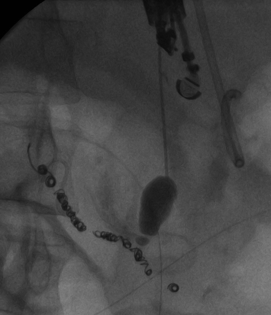
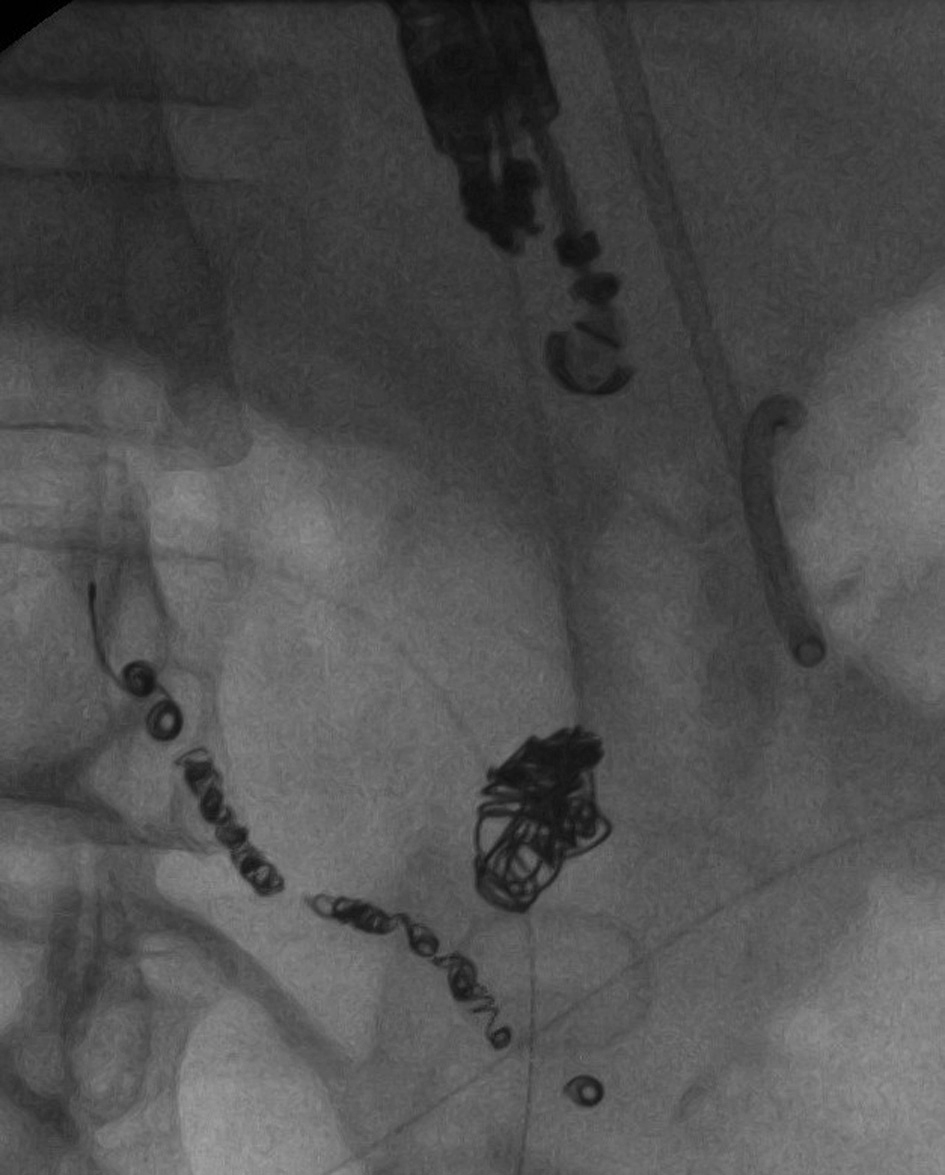
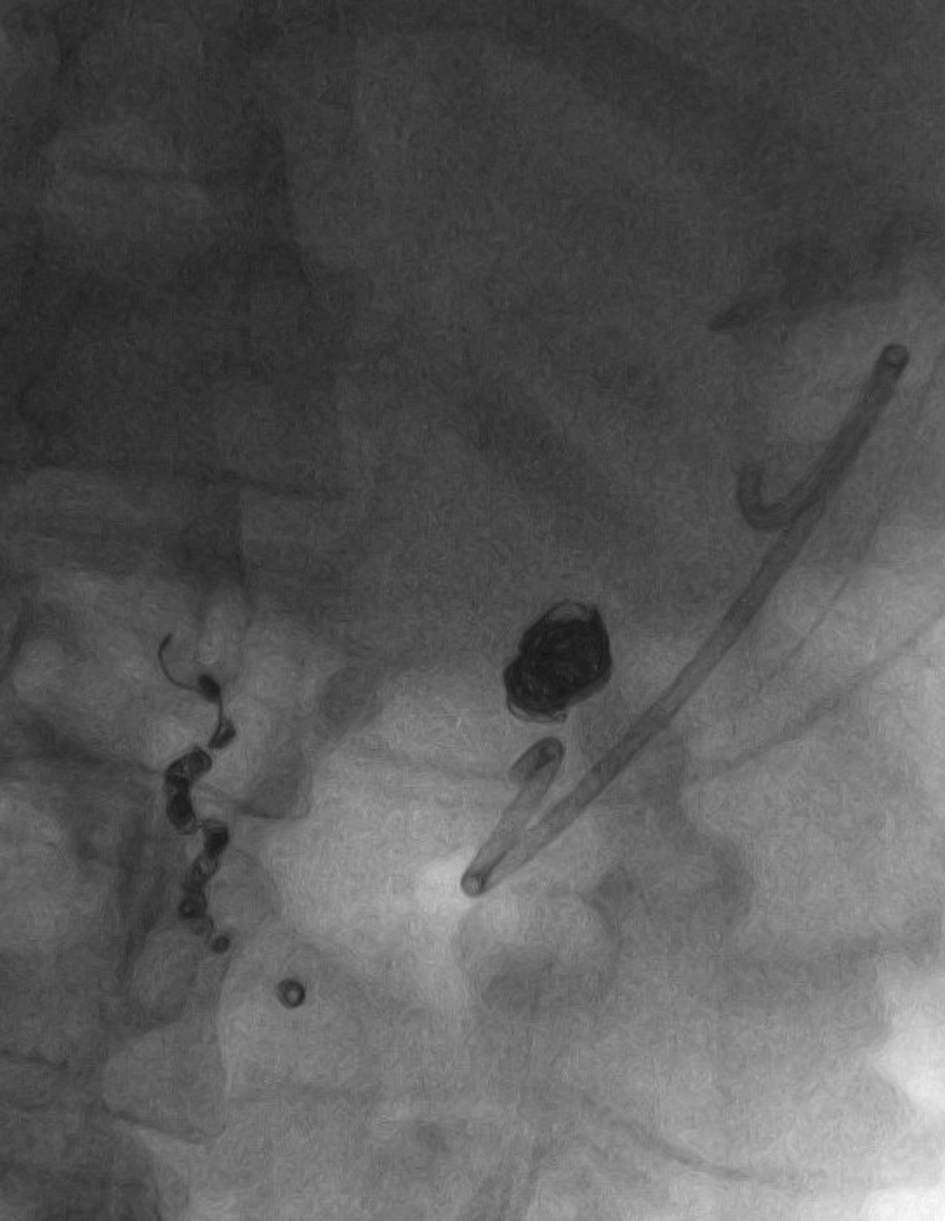
Figures