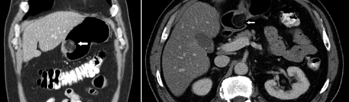
Figure 1. Axial and sagittal view of CT scan abdomen and pelvis showing a polypoid mass (white arrows) arising from gastric antrum.
| Gastroenterology Research, ISSN 1918-2805 print, 1918-2813 online, Open Access |
| Article copyright, the authors; Journal compilation copyright, Gastroenterol Res and Elmer Press Inc |
| Journal website http://www.gastrores.org |
Case Report
Volume 5, Number 5, October 2012, pages 200-204
Gastric Angiomyolipoma, a Very Rare Cause of Upper Gastrointestinal Bleeding: A Case Report and a Brief Review of Literature
Figures