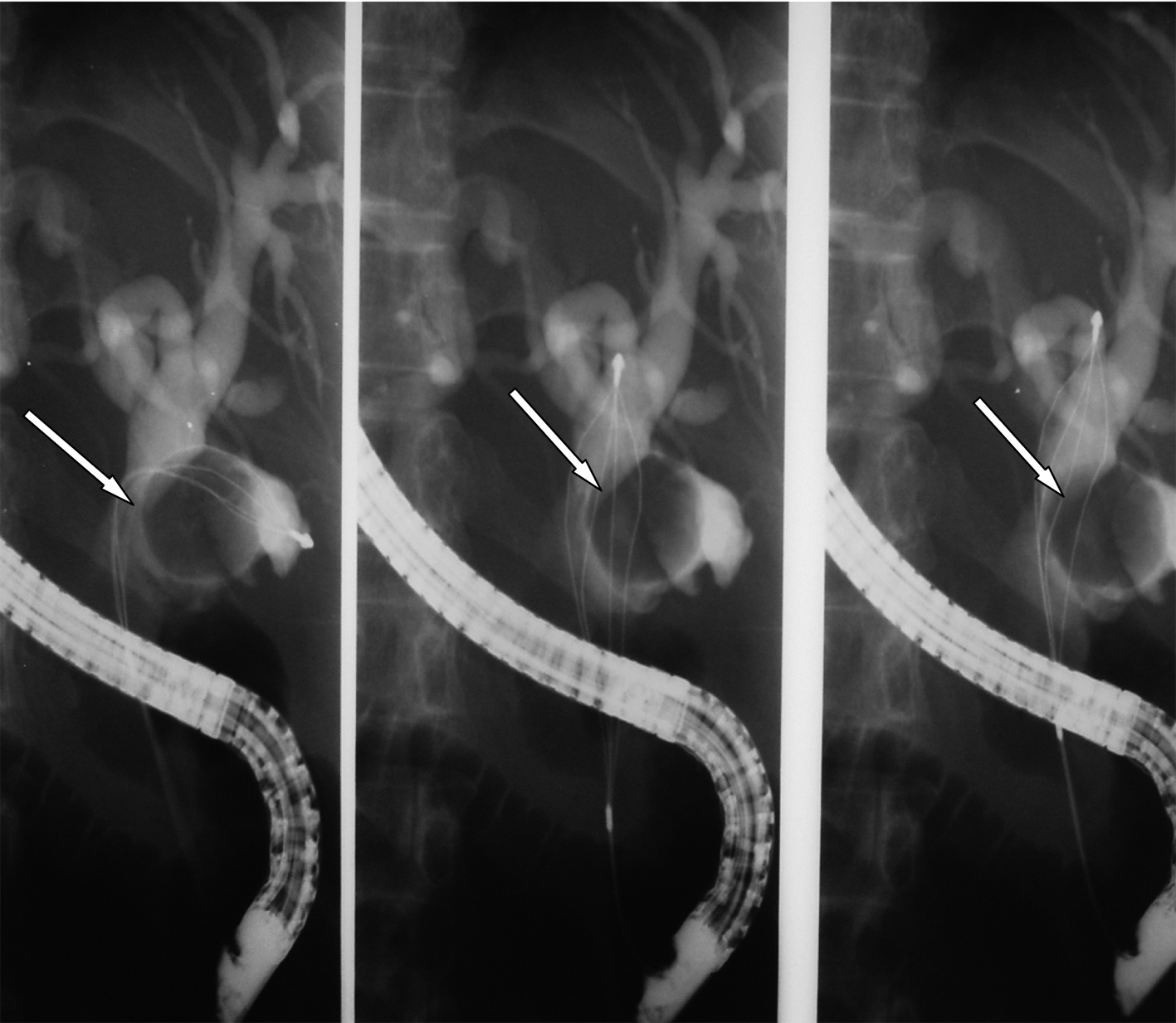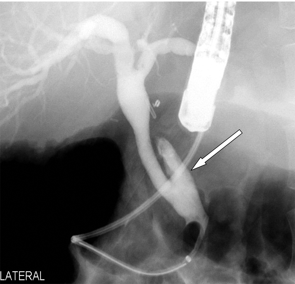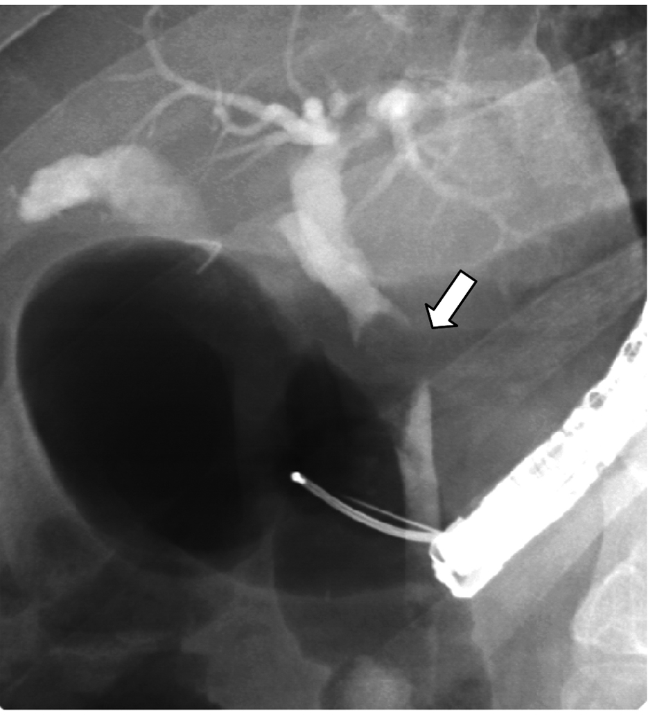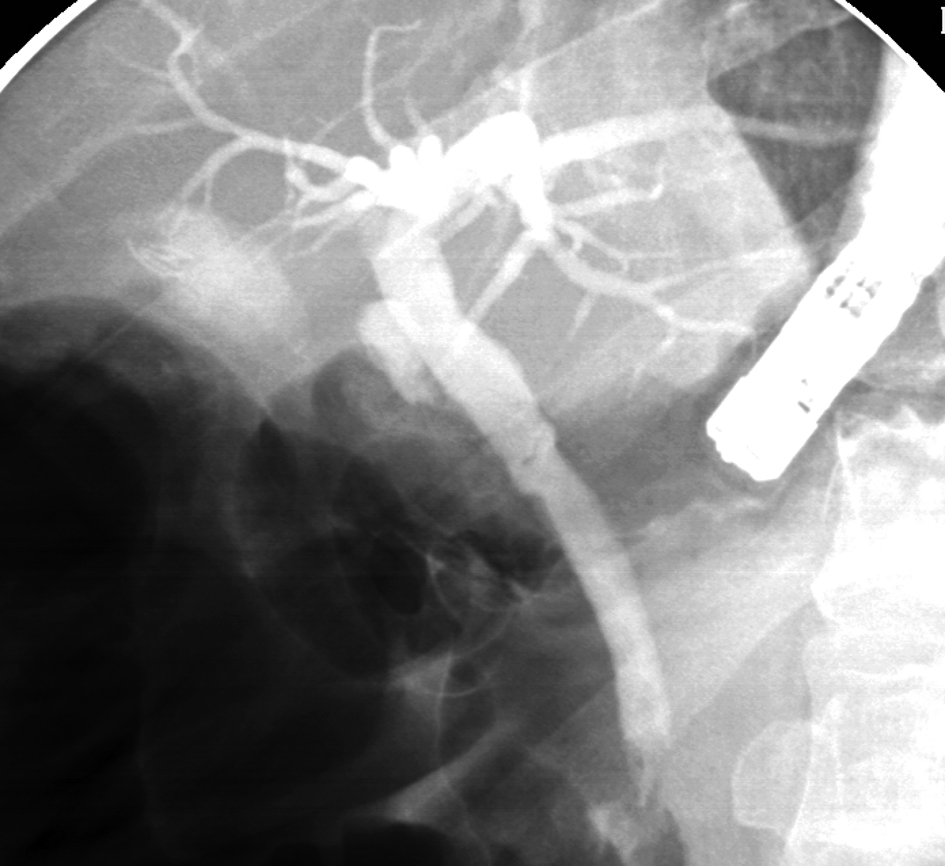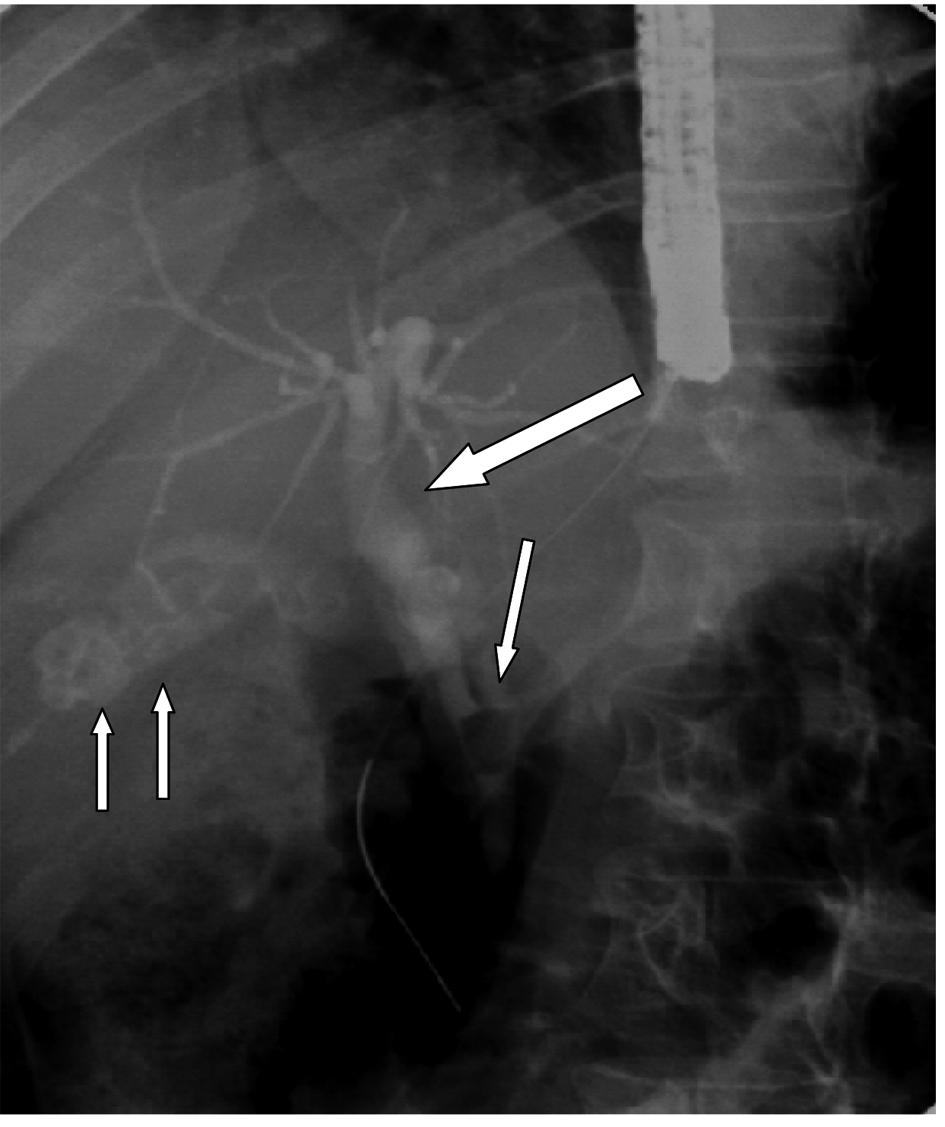
Figure 1. ERCP showing gallstones (two arrows), a long cystic duct and cystic duct stone causing Mirrizi’s syndrome (single arrow). Note the dilated common bile duct proximally (thick arrow).
| Gastroenterology Research, ISSN 1918-2805 print, 1918-2813 online, Open Access |
| Article copyright, the authors; Journal compilation copyright, Gastroenterol Res and Elmer Press Inc |
| Journal website http://www.gastrores.org |
Case Report
Volume 5, Number 4, August 2012, pages 162-166
Successful Treatment of Mirizzi’s Syndrome Using SpyGlass Guided Laser Lithotripsy
Figures