Gastroenterology Research, ISSN 1918-2805 print, 1918-2813 online, Open Access
Article copyright, the authors; Journal compilation copyright, Gastroenterol Res and Elmer Press Inc
Journal website http://www.gastrores.org

Original Article

Volume 4, Number 5, October 2011, pages 210-215


Efficacy of Narrow Band Imaging System Combined With Magnifying Endoscopy for Differentiating Type IIa Early Gastric Cancer From Adenoma

Figures

Figure 1.
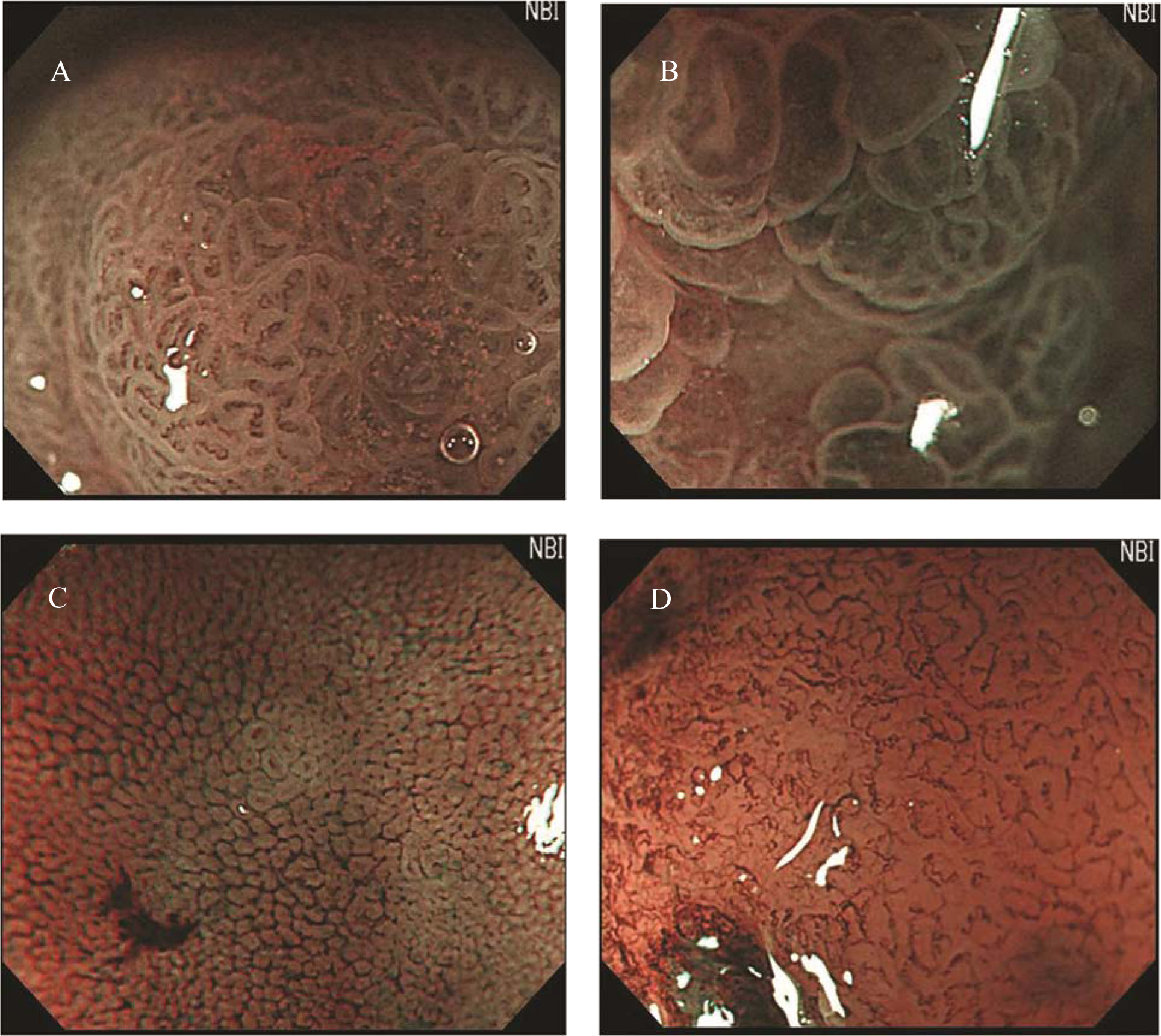
Figure 1. Classification of irregular microvascular patterns observed by MENBI: (A) Slight ISIMVPs, defined by the presence of tiny spiral blood vessels within the fine lobular superficial structure; (B) Severe ISIMVPs, defined by the presence of vertical spiral blood vessels within the coarse lobular superficial structure; (C) Fine networks, defined by fine tubular structures surrounded by a thin microvasculature; (D) Corkscrew patterns, defined by obliterated surface structures and irregular vascular patterns without loop formation. (MENBI: narrow band imaging system in combination with a magnifying endoscope; ISIMVPs: intrastructual irregular microvascular patterns)
Figure 2.
Figure 2. Image of the presence of corkscrews. (The presence of corkscrews occupying over 10% of the observed field is demonstrated (black arrows). Corkscrew was defined by obliterated surface structures and irregular vascular patterns without loop formation)

Tables

Table 1. Gross Appearance of 0-IIa Lesions
 
Adenoma (n = 32)EGC IIa (n = 14)P value
EGC IIa: type IIa early gastric cancer.
All analyses were performed by the 2-sided Fisher’s exact test except tumor size (Student t-test).
*Sensitivity 85.7%, specificity 87.5%, and positive predictive value 75.0% (for EGC IIa).
Size, mean ± SD, cm1.96 ± 0.802.02 ± 0.530.892
Reddish color, n (%)3 (9)2 (14)0.633
Obscure margin, n (%)1 (3)1 (7)0.521
Central concavity, n (%)*4 (13)12 (86)< 0.001


Table 2. Irregular Microvascular Patterns in 0-IIa Lesions
 
IMVPsAdenoma (n = 96)EGC IIa (n = 42)P value
IMVPs: irregular microvascular patterns; ISIMVPs: intrastructual irregular microvascular patterns; EGC IIa: type IIa early gastric cancer; N.A: not available. All analyses were performed by the 2-sided Fisher’s exact test.
*Sensitivity 84.3%, specificity 76.2%, and positive predictive value 89.0% (for adenoma).
Sensitivity 30.9%, specificity 93.5%, and positive predictive value 68.4% (for EGC IIa).
Sensitivity 45.2%, specificity 90.6%, and positive predictive value 67.8% (for EGC IIa).
Slight ISIMVPs*, n (%)81 (84)10 (24)< 0.001
Severe ISIMVPs†, n (%)6 (6)13 (31)< 0.001
Fine network‡, n (%)9 (10)19 (45)< 0.001
Corkscrews, n (%)0 (0)0 (0)N.A


Table 3. The Presence of Corkscrews in 0-IIa Lesions
 
The presence of corkscrewsAdenoma (n = 96)EGC IIa (n = 42)P value
EGC IIa: type IIa early gastric cancer.
The definition of the presence of corkscrew is the observation of corkscrew vascular changes in over 25% of a selected field.
All analyses were performed by the 2-sided Fisher’s exact test.
*Sensitivity 9.5%, specificity 100%, and positive predictive value 100% (for EGC IIa).
Positive, n (%)*0 (0)4 (9.5)0.008
Negative, n (%)96 (100)38 (88)


Table 4. The Presence of Corkscrews and IMVPs
 
IMVPsThe presence of corkscrewsP value
PositiveNegative
IMVPs: irregular microvascular patterns; ISIMVPs: intrastructual irregular microvascular patterns; EGC IIa: type IIa early gastric cancer.
All analyses were performed by the 2-sided Fisher’s exact test.
Slight ISIMVPs, n = 91 (%)0 (0)91 (100)0.012
Severe ISIMVPs, n = 19 (%)4 (21)15 (79)< 0.001
Fine network, n = 28 (%)0 (0)28 (0)0.582