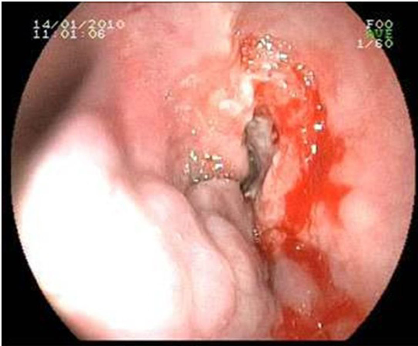
Figure 1. Endoscopic appearance of the esophageal mass (right) and the varices (left side).
| Gastroenterology Research, ISSN 1918-2805 print, 1918-2813 online, Open Access |
| Article copyright, the authors; Journal compilation copyright, Gastroenterol Res and Elmer Press Inc |
| Journal website http://www.gastrores.org |
Case Report
Volume 4, Number 2, April 2011, pages 84-87
Synchronous Esophageal Squamous Cell Carcinoma and Esophageal Variceal Bleeding due to Idiopathic Portal Hypertension: A Case Report
Figures