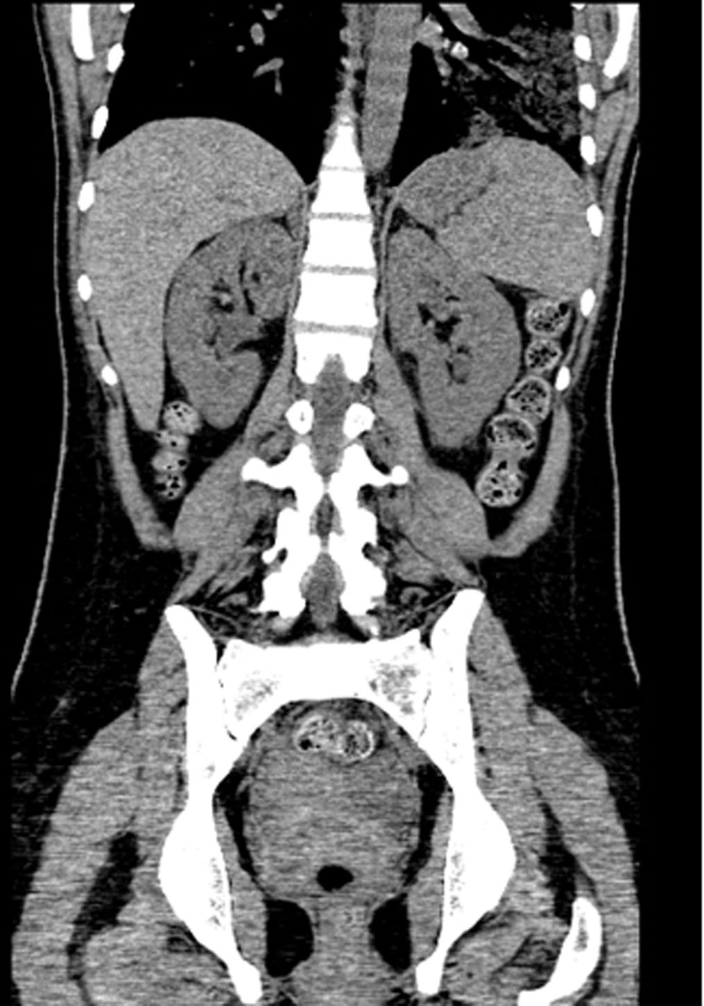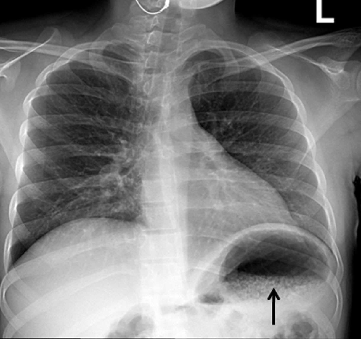
Figure 1. Chest roentgenogram showing innumerable bubbles outlining the stomach in a mottled distribution (arrow).
| Gastroenterology Research, ISSN 1918-2805 print, 1918-2813 online, Open Access |
| Article copyright, the authors; Journal compilation copyright, Gastroenterol Res and Elmer Press Inc |
| Journal website http://www.gastrores.org |
Case Report
Volume 4, Number 2, April 2011, pages 76-79
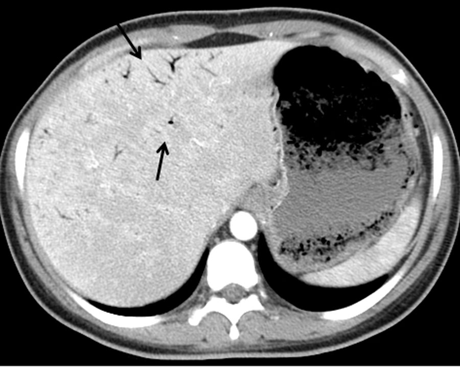
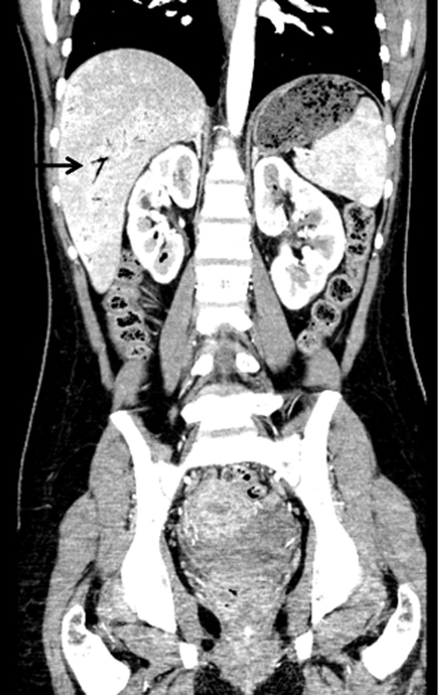
Emphysematous Pyelonephritis Associated With Emphysematous Gastritis and Air in the Portal Vein
Figures