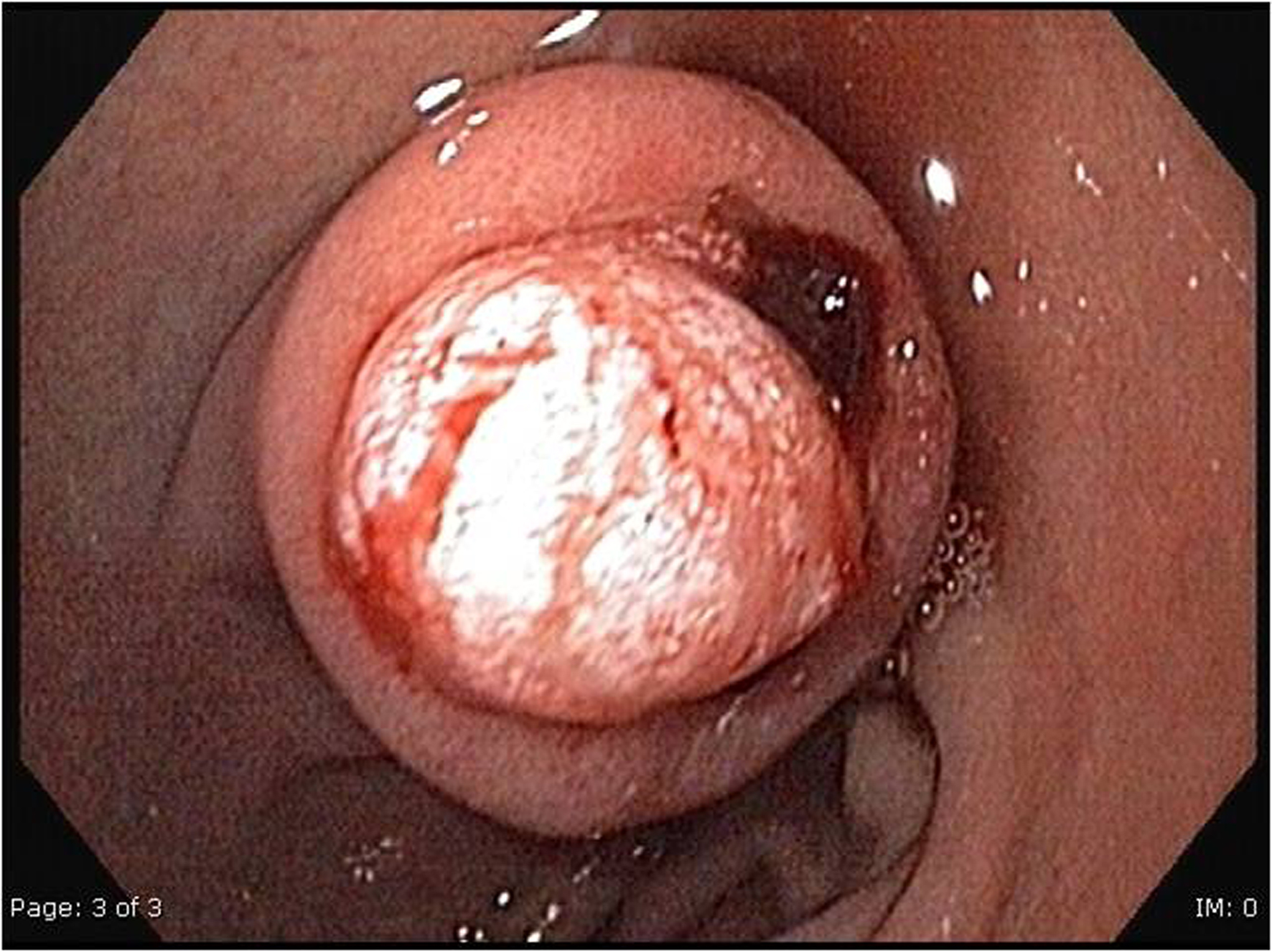
Figure 1. Gastroscopy shows a submucosal tumor of the bulbus duodeni of 1.0 x 2.0 cm in diameter, with a central bleeding stigma.
| Gastroenterology Research, ISSN 1918-2805 print, 1918-2813 online, Open Access |
| Article copyright, the authors; Journal compilation copyright, Gastroenterol Res and Elmer Press Inc |
| Journal website http://www.gastrores.org |
Case Report
Volume 3, Number 6, December 2010, pages 290-292
Duodenal Lipoma as a Rare Cause of Upper Gastrointestinal Bleeding
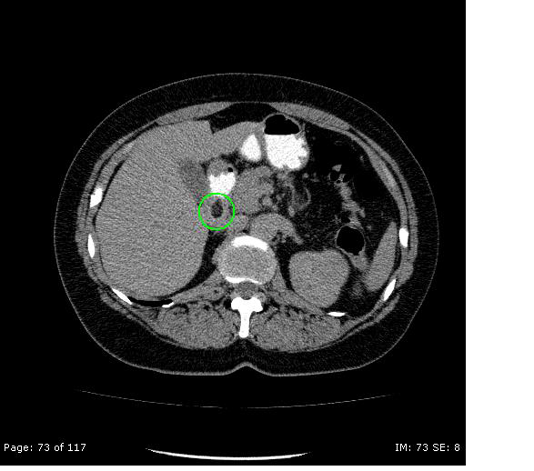
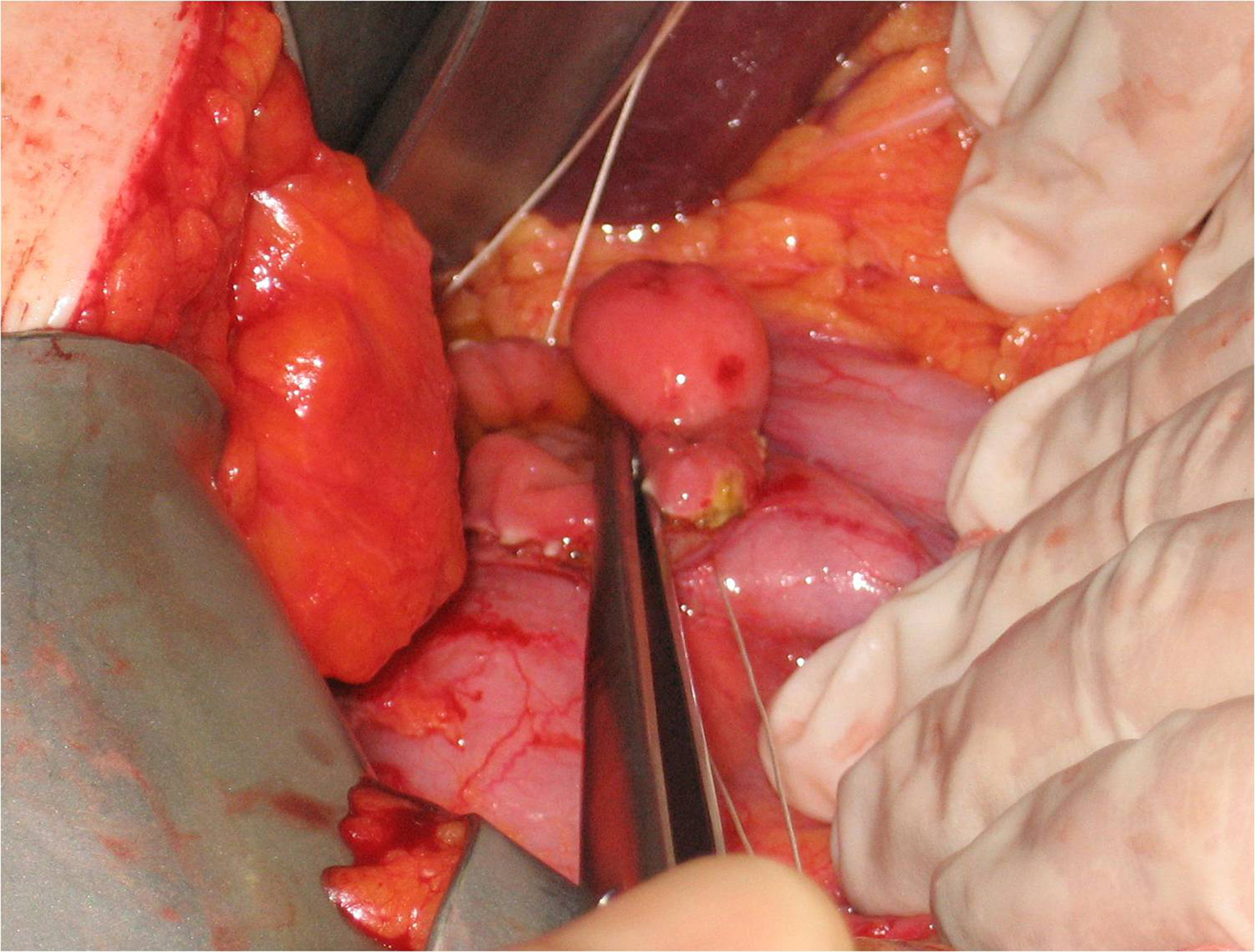
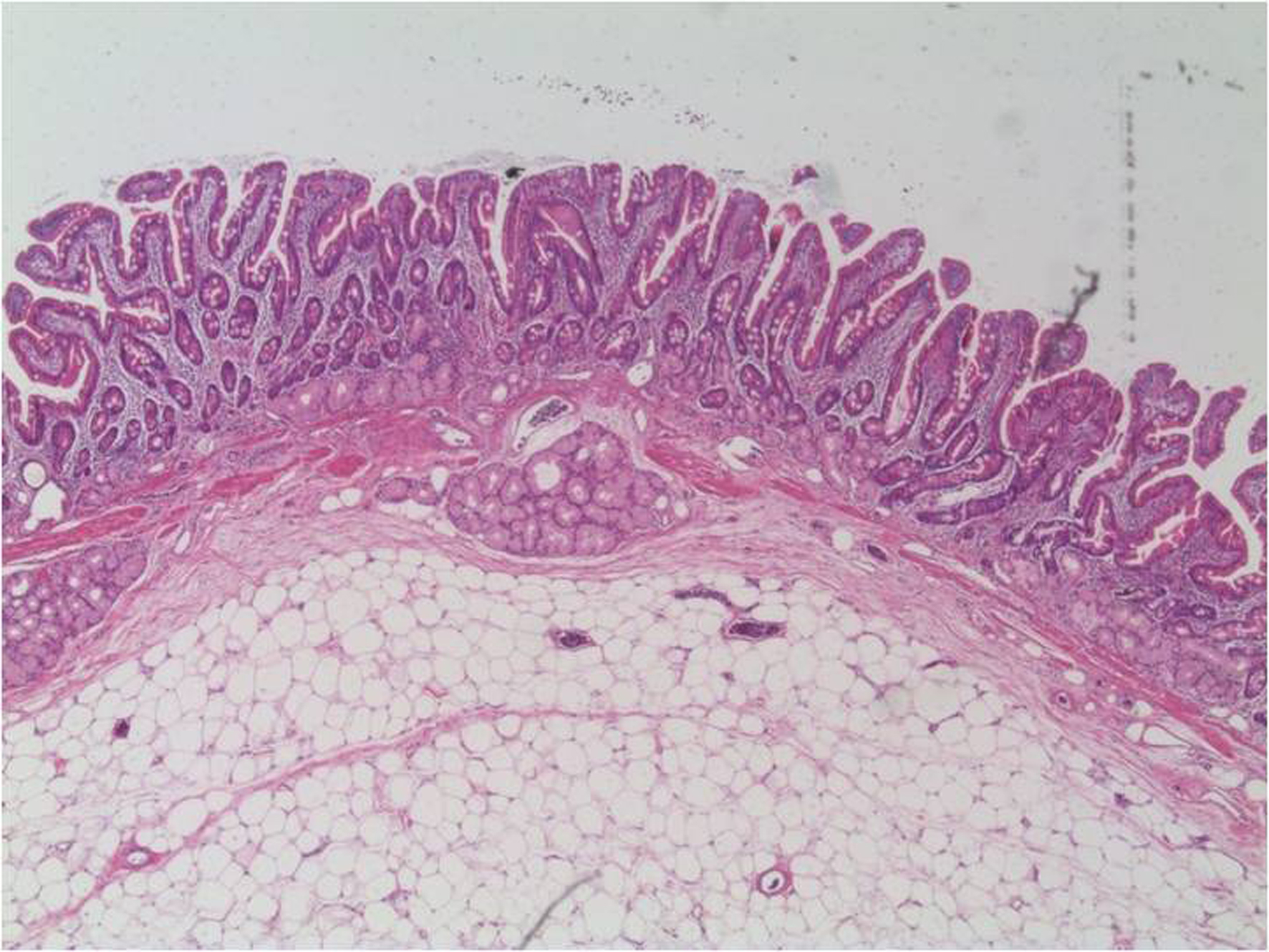
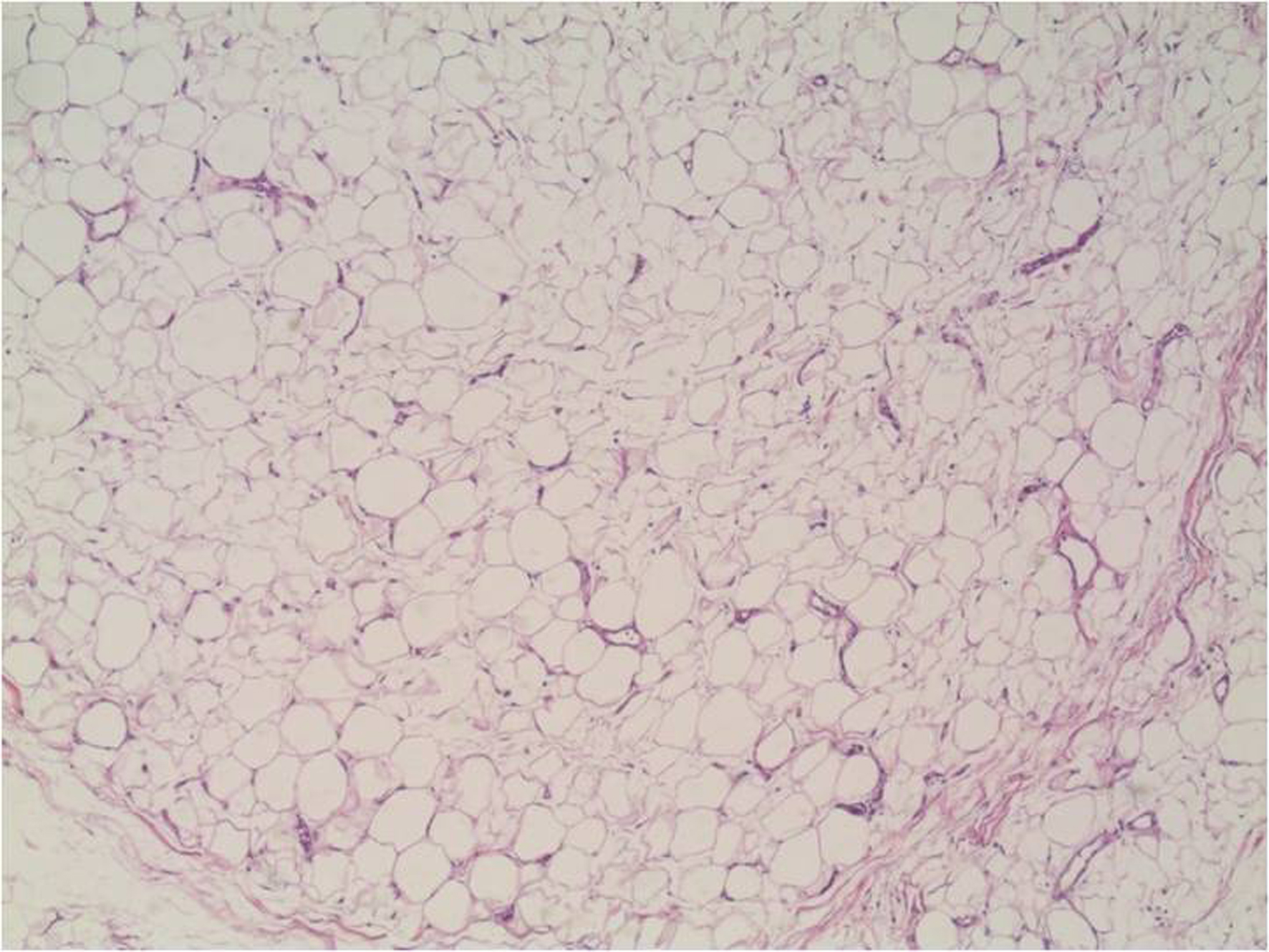
Figures