Gastroenterology Research, ISSN 1918-2805 print, 1918-2813 online, Open Access
Article copyright, the authors; Journal compilation copyright, Gastroenterol Res and Elmer Press Inc
Journal website http://www.gastrores.org

Case Report

Volume 3, Number 6, December 2010, pages 276-280


Gastric Schwannoma Presenting as an Incidentaloma on CT-Scan and MRI

Figures

Figure 1.
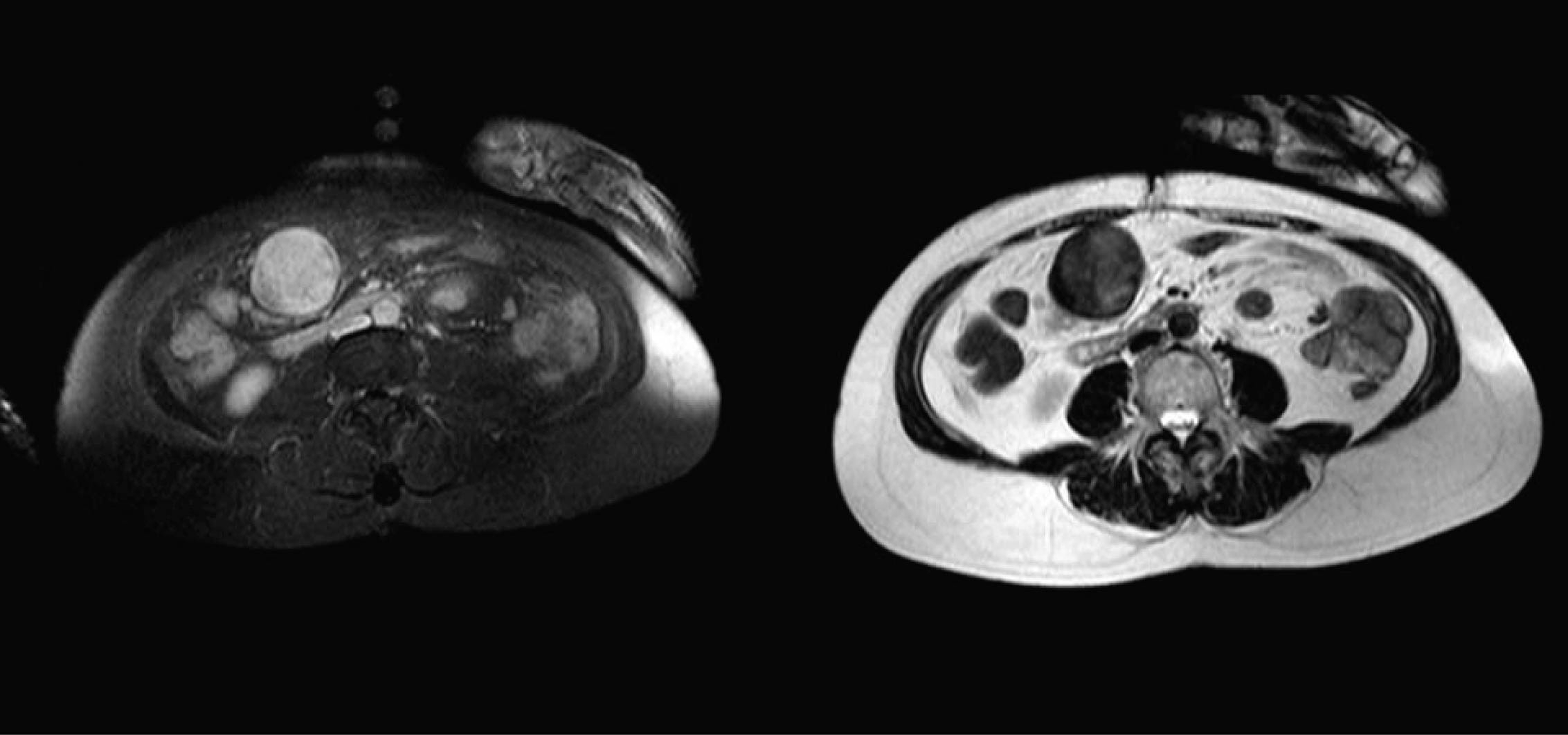
Figure 1. Left a contrast enhanced T1 weighted image with an overall inhomogeneous high signal pattern; Right a T2 weighted image with a low to intermediate signal pattern.
Figure 2.
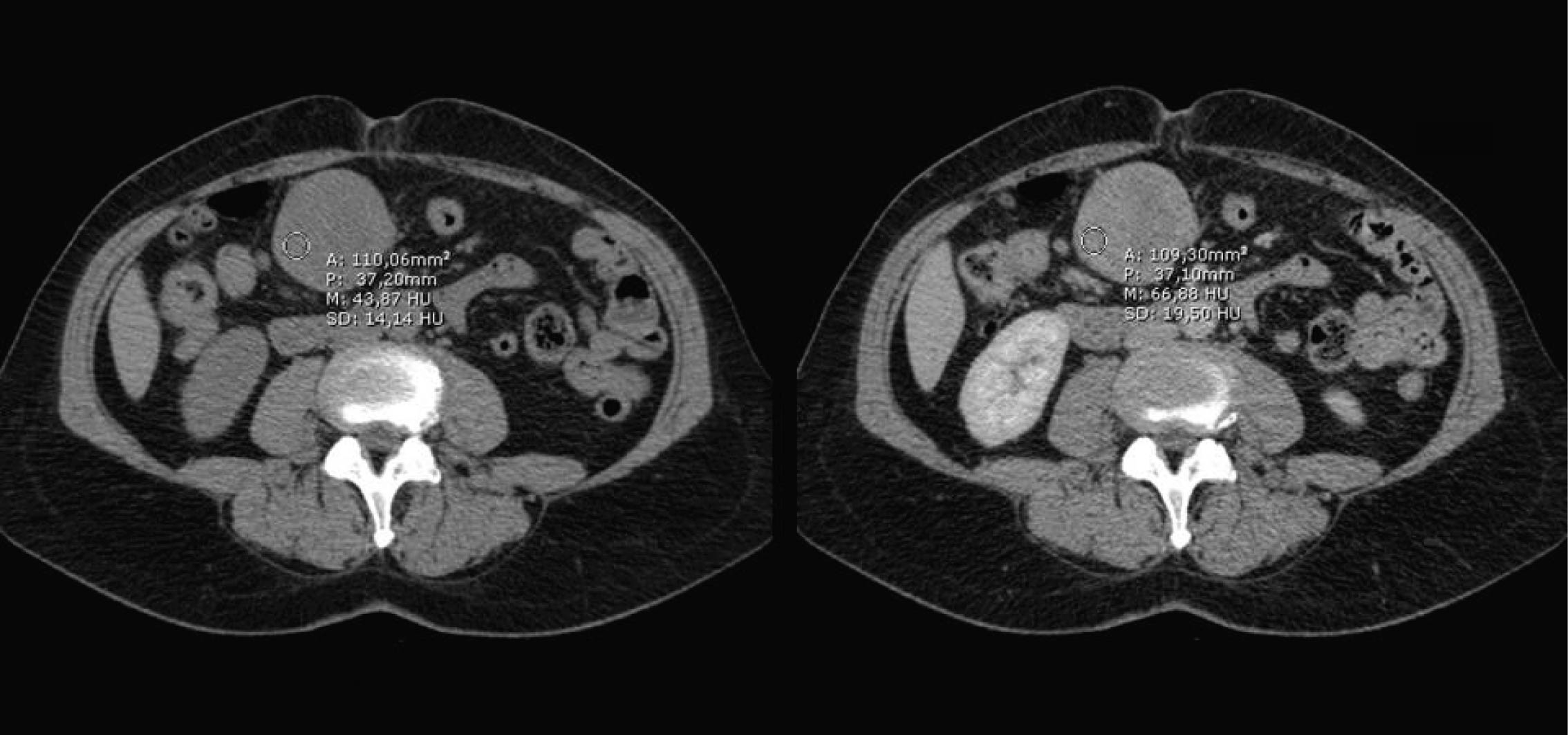
Figure 2. CT-scan showing on the left a non contrast image of the lesion and on the right a post contrast image showing slight enhancement, depicted as the drawn circle with the mean Hounsfield units.
Figure 3.
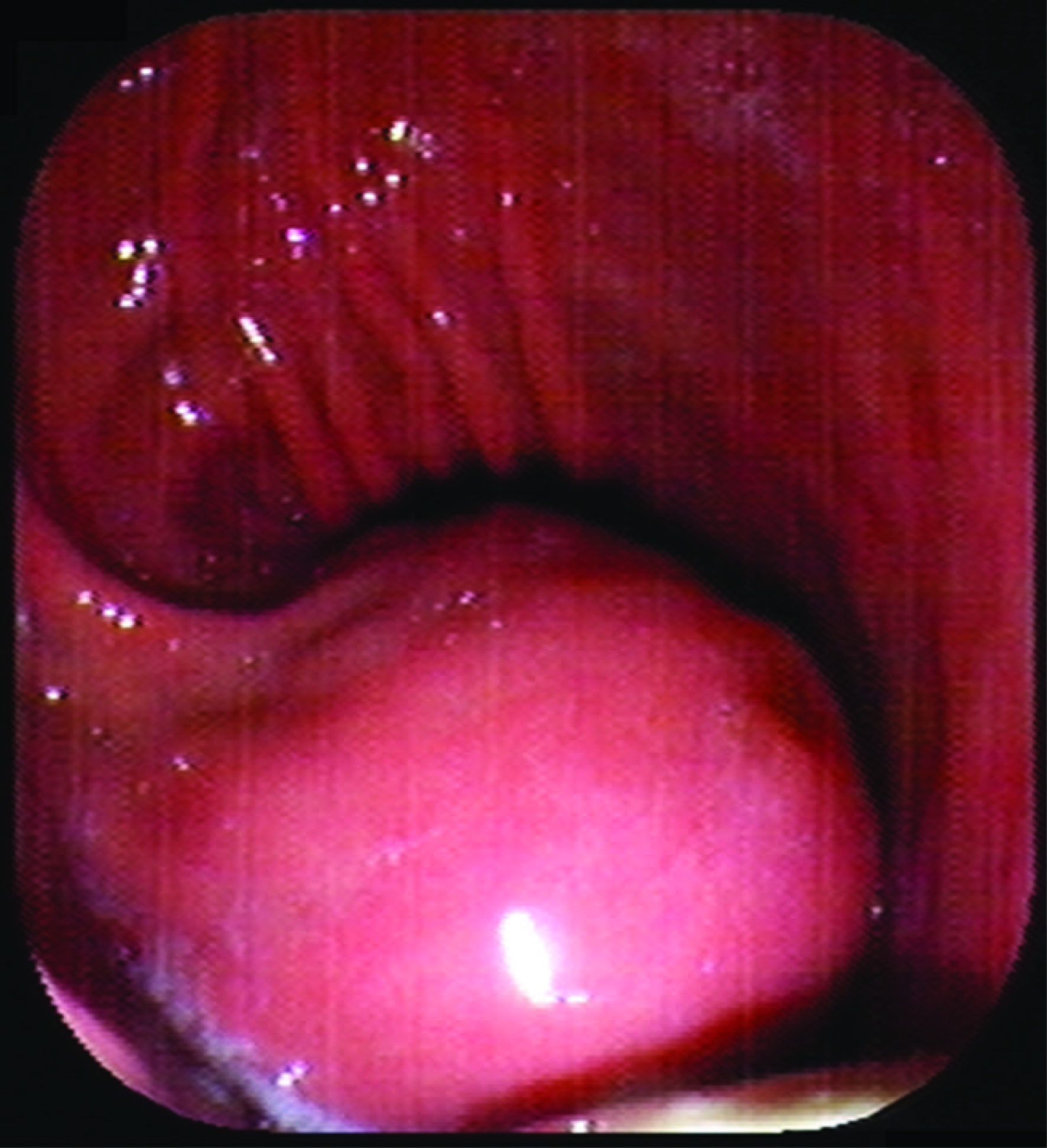
Figure 3. An exophytically growing, submucosal mass is seen in the stomach during endoscopy.
Figure 4.
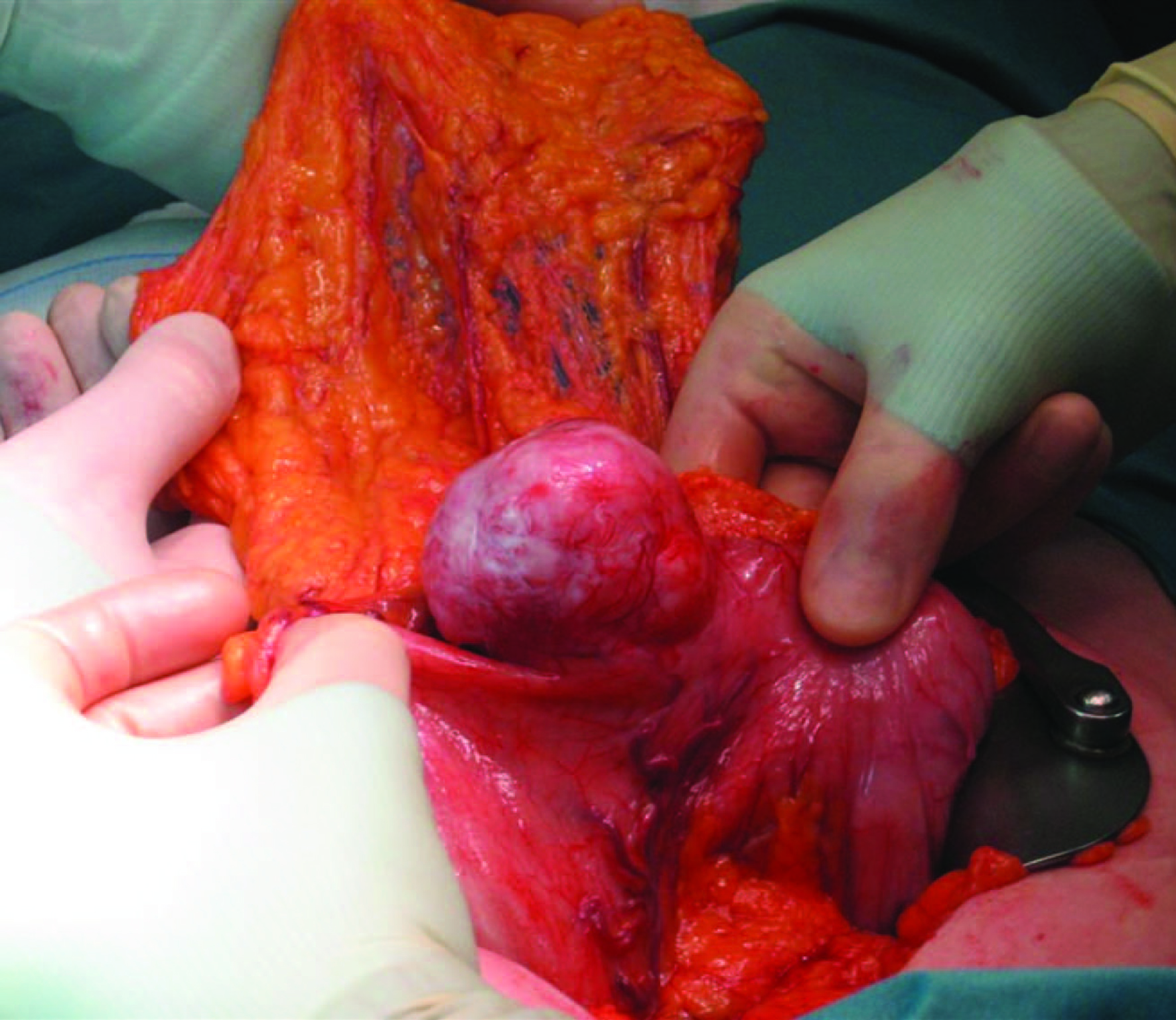
Figure 4. Macroscopic inspection during surgery showed a tumor with a diameter of 7 cm in the distal stomach.
Figure 5.
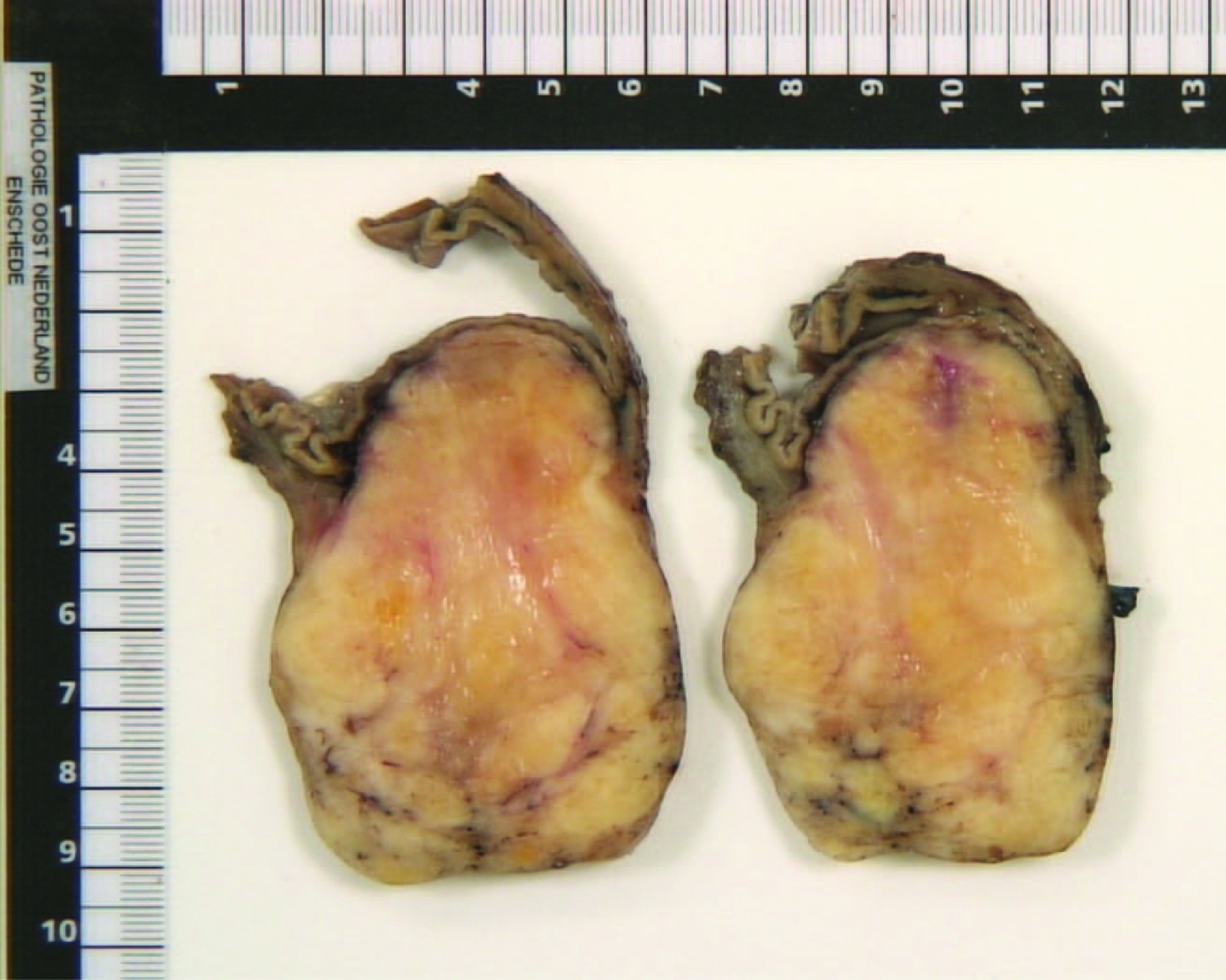
Figure 5. Cross section of the tumor, revealing an off-white mass with an intact overlying mucosal layer.
Figure 6.
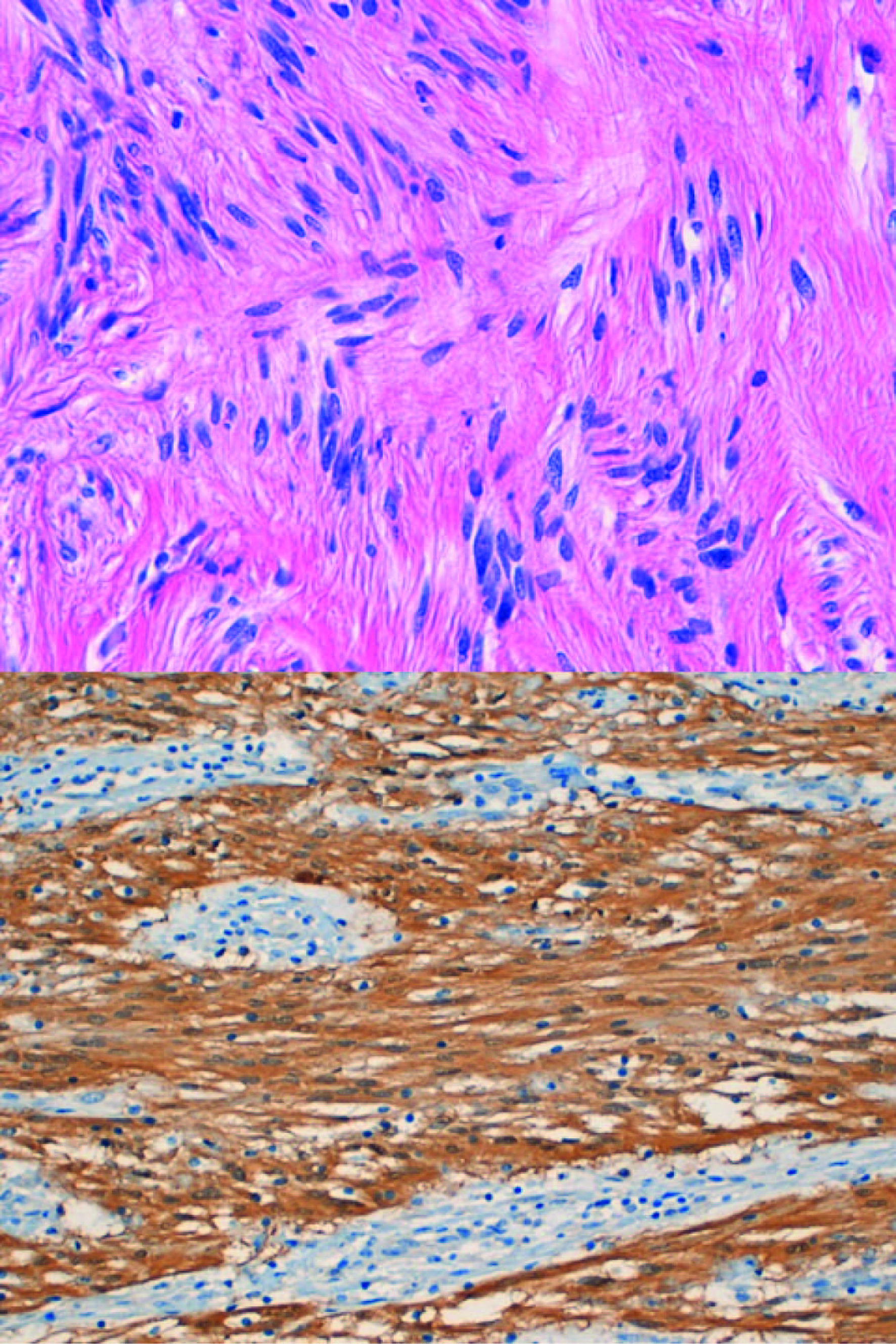
Figure 6. Upper: Schwannoma (Spindle cells with nuclear palisading); Lower: diffuse S100 positivity in Schwann cells.
Figure 7.
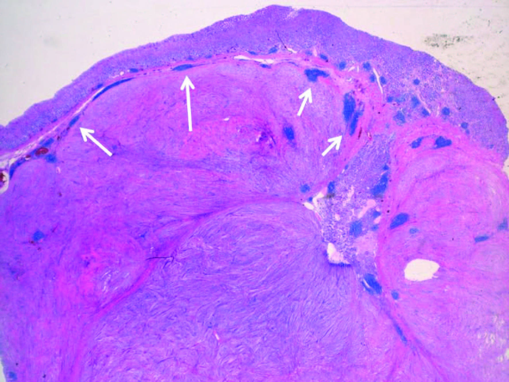
Figure 7. Lymphoid infiltrates (see arrows) at the periphery of the lesion are a common and characteristic feature.