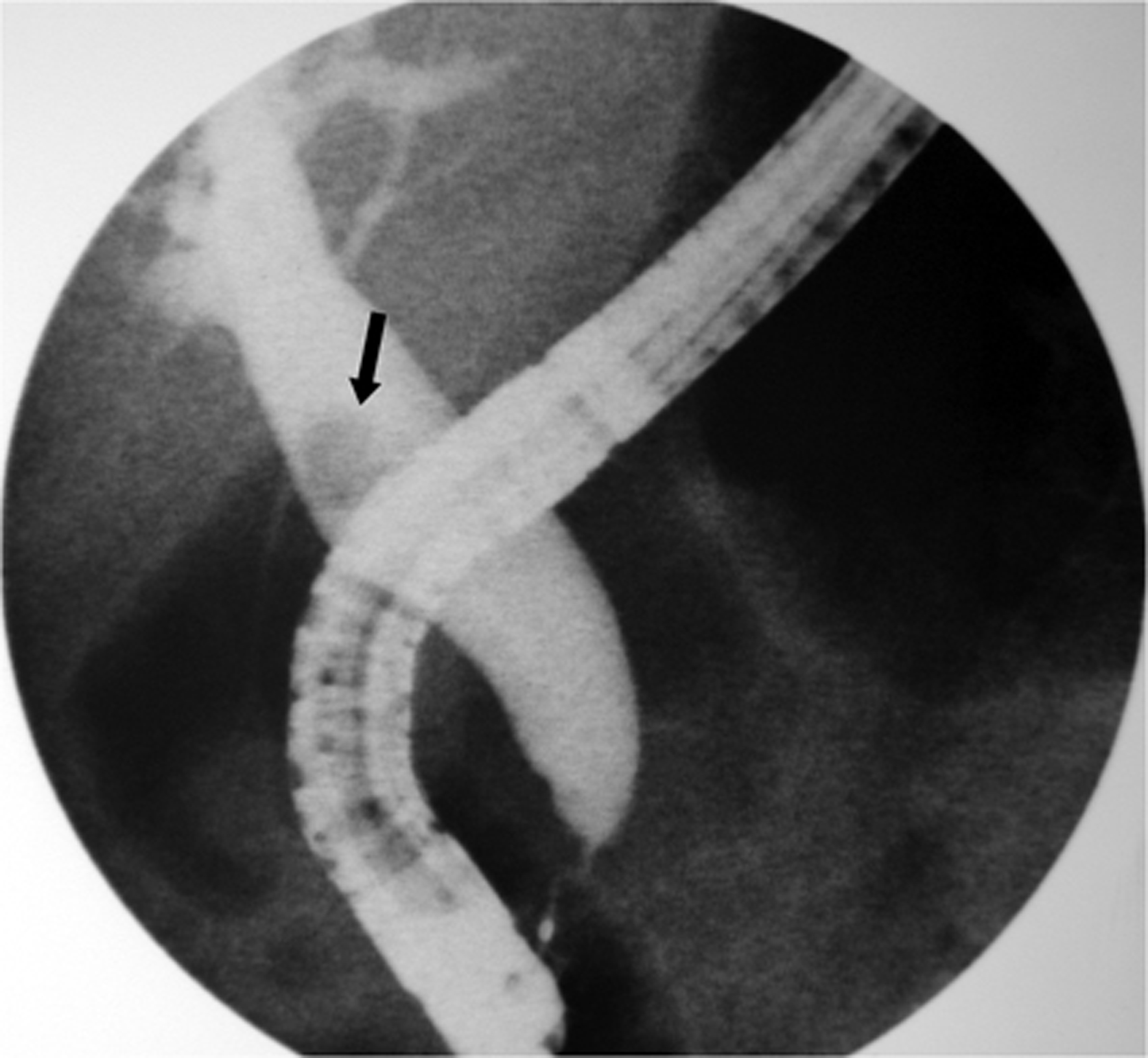
Figure 1. Cholangiography showing a dilatated common bile duct with a single stone (black arrow).
| Gastroenterology Research, ISSN 1918-2805 print, 1918-2813 online, Open Access |
| Article copyright, the authors; Journal compilation copyright, Gastroenterol Res and Elmer Press Inc |
| Journal website http://www.gastrores.org |
Case Report
Volume 3, Number 5, October 2010, pages 216-218
Subcutaneous Emphysema, Pneumothorax and Pneumomediastinum Following Endoscopic Sphincterotomy
Figures