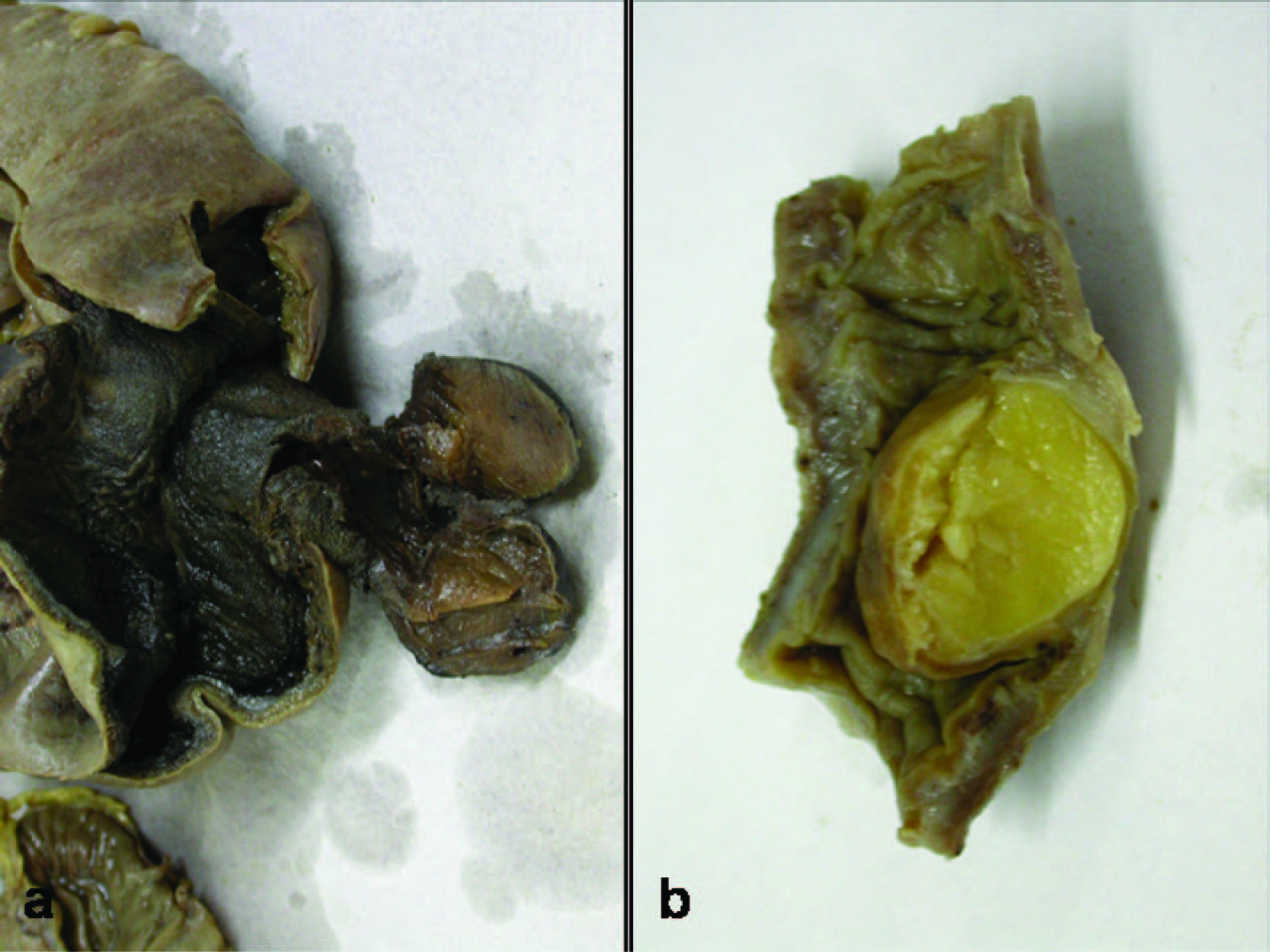
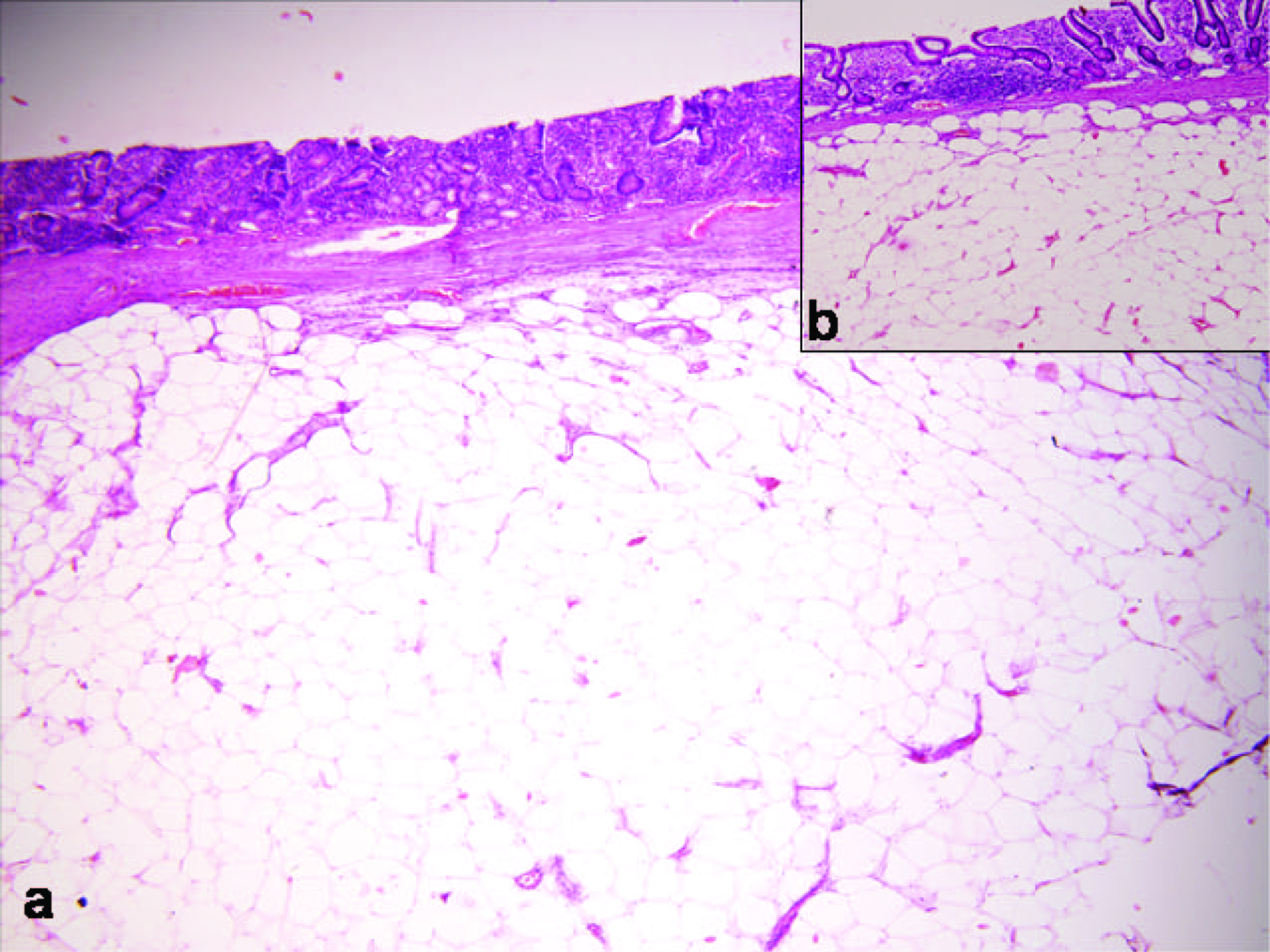
Figure 1. (a) Gangrene intestine with sesile Lipomatous polyp; (b) Submucosal Lipomatous polyp.
| Gastroenterology Research, ISSN 1918-2805 print, 1918-2813 online, Open Access |
| Article copyright, the authors; Journal compilation copyright, Gastroenterol Res and Elmer Press Inc |
| Journal website http://www.gastrores.org |
Case Report
Volume 3, Number 5, October 2010, pages 229-231
Lipomatous Polyp Presenting With Intestinal Intussusception in Adults: Report of Four Cases
Figures
Table
| Age/Sex | Presentation | Location | Gross | Microscopy | |
|---|---|---|---|---|---|
| Case 1 | 45/M | Pain, Abdominal distension, non passage of stools, flatus | Ileum, 14 cm from ileocolic junction | Ileoileal intussusseption; A polyp measuring 2 cm at the lading end of intussusceptions; 2 small proximal perforations | Submucosal Lipomatous polyp; Microscopic evidence of perforation |
| Case 2 | 55/M | Recurrent vomiting, abdominal distension | Ileum, 16 cm from ileocolic junction | Ileocolic Intussusceptions; Gangrene of the bowel loops; Small polyp 1 cm diameter | Gangrenous intestine; Lipomatous polyp of the small intestine; Microscopic evidence of perforation |
| Case 3 | 50/M | Vomiting abdominal distension, inability to pass stools for 10 days | 24 cm from the Ileocolic junction, proximal perforation | Ileoileal intussusseption polyp 2.5 cm diameter | Gangrenous intestine; Lipomatous polyp of the small intestine; Mesentric artery thrombosis |
| Case 4 | 66/M | Sub acute intestinal obstruction | Ascending colon, 5 cm from Ileocolic junction | Colocolic intussusception; Polyp 3 cm diameter | Colonic Submucosal; Lipomatous polyp |