Gastroenterology Research, ISSN 1918-2805 print, 1918-2813 online, Open Access
Article copyright, the authors; Journal compilation copyright, Gastroenterol Res and Elmer Press Inc
Journal website http://www.gastrores.org

Case Report

Volume 2, Number 2, April 2009, pages 122-125


Ileal Varices Treated with Balloon-Occluded Retrograde Transvenous Obliteration

Figures

Figure 1.
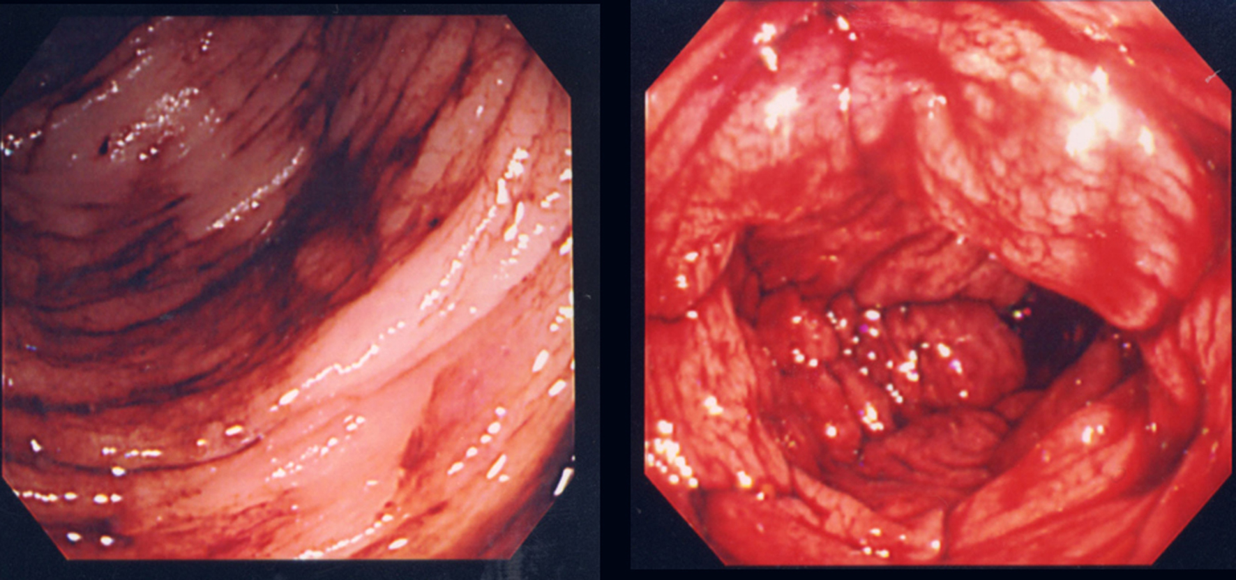
Figure 1. Colonoscopy revealed blood retention in the entire colon, but no bleeding lesion was detected.
Figure 2.
Figure 2. Computed tomography images of a vessel in the ileum (arrow) and its connection to the right testicular vein (arrowhead).
Figure 3.
Figure 3. (a) Ileal varices (arrowhead) communicating with the right testicular vein (arrow) were found using retrograde transvenous venography. (b) Balloon occluded retrograde transvenous obliteration for ileal varices (arrowhead) was performed via the efferent vein of the varices.
Figure 4.
Figure 4. Following balloon occluded retrograde transvenous obliteration, both ileal varices (arrow) and the afferent vein were not visible by venography the next day.
Figure 5.
Figure 5. (a) Computed tomography image of ileal vessels before balloon occluded retrograde transvenous obliteration. (b) The ileal vessels are no longer visible following balloon occluded retrograde transvenous obliteration.