Gastroenterology Research, ISSN 1918-2805 print, 1918-2813 online, Open Access
Article copyright, the authors; Journal compilation copyright, Gastroenterol Res and Elmer Press Inc
Journal website http://www.gastrores.org

Original Article

Volume 2, Number 2, April 2009, pages 100-103


Benign Tumors of the Esophagus: A Histopathologic Study of 49 Cases among 931 Consecutive Esophageal Biopsies

Figures

Figure 1.
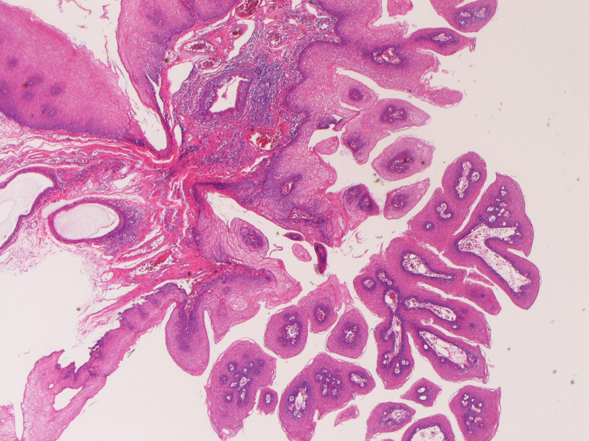
Figure 1. Squamous papilloma of the esophagus. Papillary proliferation of mature squamous epithelium is seen. HE, x20
Figure 2.
Figure 2. Granular cell tumor of the esophagus. A: Histologic features. Large cells with acidophilic granular cytoplasm are seen. Many hyaline globules are also seen. HE, x200 B: The cytoplasm and hyaline globules are positive with PAS stain. PAS, x200 C: The tumor cells are positive for S100 protein. Immunostaining, x200 D: The tumor cells are positive for neuron-specific enolase. Immunostaining, x200.
Figure 3.
Figure 3. Tubular adenoma of the esophagus. A: adenomatous tubules are seen. HE, x200 B: The tumor cells are very focally positive for p53 protein. Immunostaining, x100 C: The Ki-67 labeling is 26%. Immunostaining (MIB1), x200
Figure 4.
Figure 4. Leiomyoma of the esophagus. A: Low power view shows well-define nodule composed of acidophilic spindle cells. HE, x40 B: The tumor is composed of spindle cells considered as smooth muscle cells. HE, x200