Gastroenterology Research, ISSN 1918-2805 print, 1918-2813 online, Open Access
Article copyright, the authors; Journal compilation copyright, Gastroenterol Res and Elmer Press Inc
Journal website http://www.gastrores.org

Original Article

Volume 2, Number 1, February 2009, pages 29-34


Combined Intestinal and Biliary Stenting in Gastric Outlet and Biliary Obstruction

Figures

Figure 1.
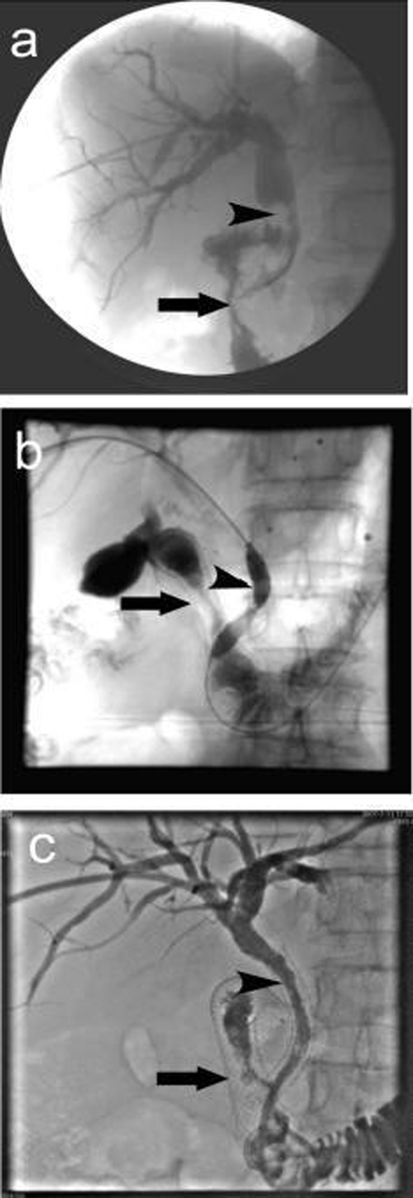
Figure 1. Procedure of double stenting. (a) Duodenal stenosis combined with common bile duct lesions. The arrow indicates descending duodenal stenosis, the arrow head shows the irregular contrast deficiency of lower common bile duct, which might be invaded by tumors which led to the expansion of common bile duct above the stenosis; (b) Biliary stent placement, the arrow shows duodenal stent, the arrow head shows balloon dilatation of common bile duct stenosis and the mesh of intestinal stent; (c) Biliary stent placement, the arrow shows duodenal stent, the arrow head shows biliary stent. The cholangiography showed both were patent.
Figure 2.
Figure 2. The relationship of the intestinal and biliary stents. (a) anteroposterior; (b) lateral.

Table

Table 1. Serum bilirubin and liver function before and 2 weeks after stenting
 
Pre-stentingPost-stenting (2 weeks)P value
N3232
TBil (µmol/L)276.54 ± 72.67116.37 ± 48.42< 0.01
DBil (µmol/L)152.32 ± 42.6465.70 ± 31.93< 0.01
ALT (U/L)109.22 ± 37.5646.77 ± 33.92< 0.01
AST (U/L)72.38 ± 13.8634.17 ± 18.20< 0.01
ALP (U/L)486.35 ± 129.67117.56 ± 93.42< 0.01
γ-GT (U/L)603.22 ± 145.79247.83 ± 63.07< 0.01