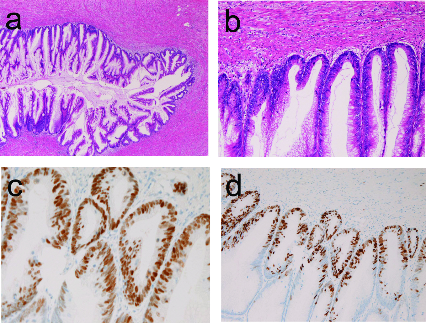
Figure 1. (a) Low power view of papillary adenocarcinoma of the appendix in case 1. Papillary proliferation is apparent. No invasion is seen. HE, x 20. (b) Higher power view of Figure 1A. The cellular atypia is evident. HE, x 200. (c) The tumor cells are positive for p53 protein, Immunostaining, x 200. (d) The Ki-67 labeling is 90%. Immunostaining, x 100.
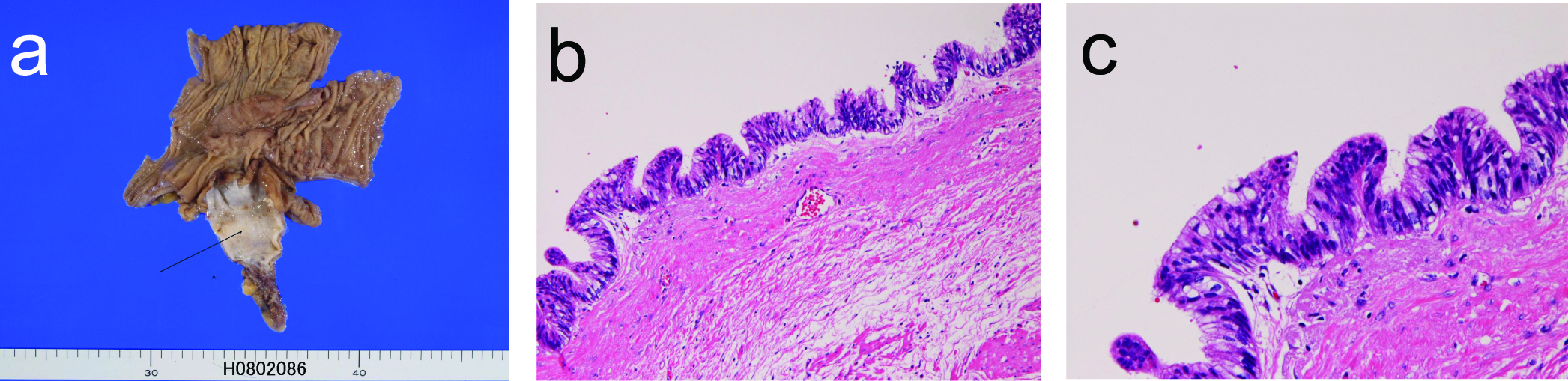
Figure 3. (a) The resected appendix shows cystic dilation (arrow) in case 3. (b) The cystic lining is adenocarcinoma cells. HE, x 100. (c) The cellular atypia is enough to be regarded as adenocarcinoma. HE, x 200.
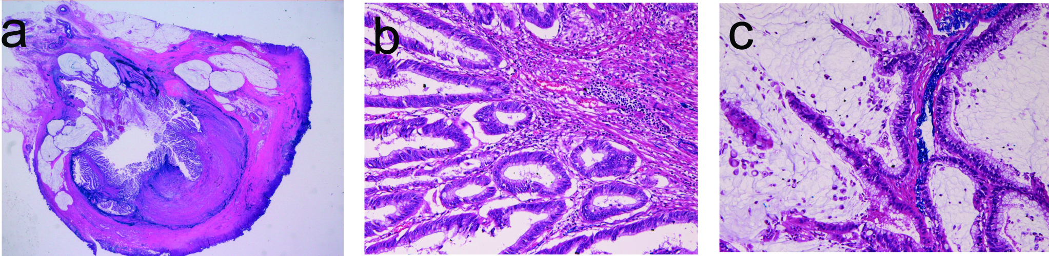
Figure 4. (a) Loupe figures of appendiceal mucinous adenocarcinoma in case 4. Papillary epithelial proliferation and intraluminal mucus are evident. No invasion of tumor cells is recognized. (b) Higher power view of the mucosa of the appendix. Papillary adenocarcinoma is evident. HE, x 200. (c) The intraluminal area show adenocarcinoma cells and much mucus. HE, x 200.