Gastroenterology Research, ISSN 1918-2805 print, 1918-2813 online, Open Access
Article copyright, the authors; Journal compilation copyright, Gastroenterol Res and Elmer Press Inc
Journal website http://www.gastrores.org

Case Report

Volume 2, Number 6, December 2009, pages 364-366


Non-functioning Well Differentiated Endocrine Carcinoma of the Pancreas

Figures

Figure 1.
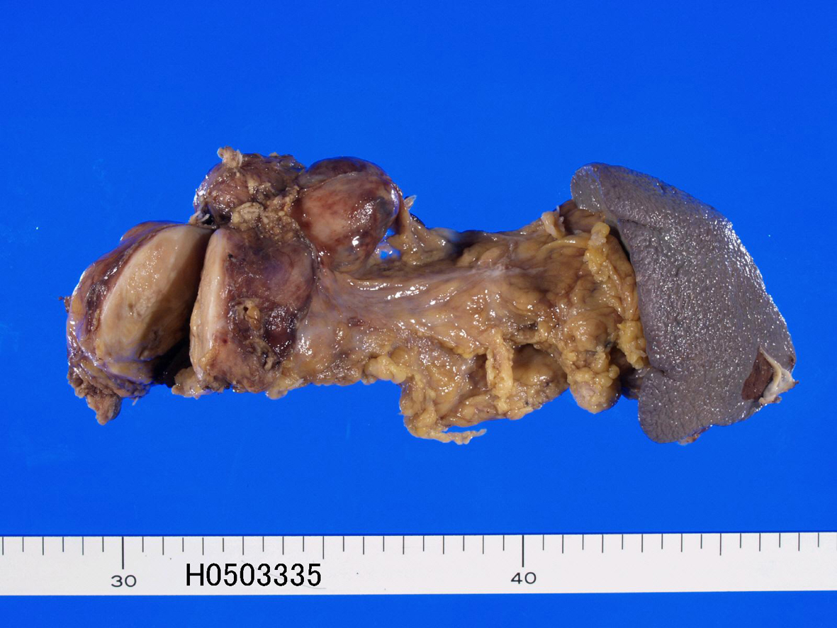
Figure 1. Gross features of resected pancreas. A well defined tumor measuring 60 x 55 x 50 mm is recognized (left). Lymph node metastasis is also seen (left upper).
Figure 2.
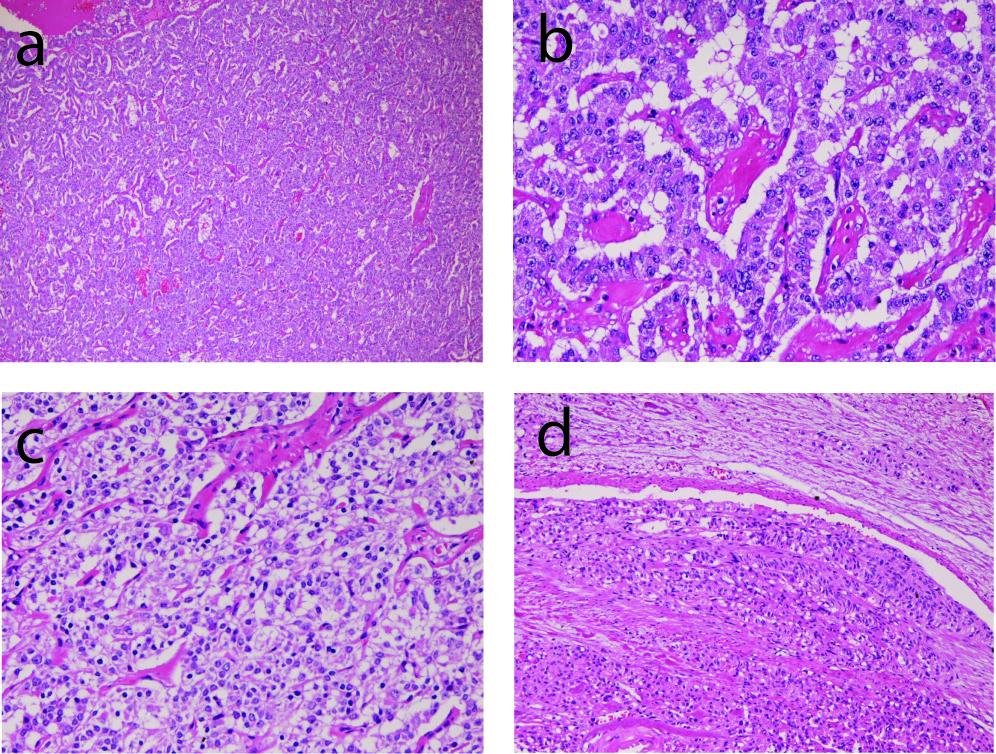
Figure 2. Microscopic findings of the tumor. (a) very low power view of the tumor. Tumor cells are arranged in a trabecular pattern. HE, x20. (b) tumor cells are arranged in a trabecular pattern. The nuclei show ‘salt and pepper’ appearances. HE, x200. (c) clear cell change of tumor cells. HE, x200. (d) vascular invasion of tumor cells. HE, x100.
Figure 3.
Figure 3. Immunohistochemical findings of the tumor. (a) tumor cells are positive for cytokeratin. Immunostaining, x200. (b) Tumor cells are positive for synaptophysin. Immunostaining, x200. (c) Tumor cells are positive for CD56. Immunostaining, x200.