Gastroenterology Research, ISSN 1918-2805 print, 1918-2813 online, Open Access
Article copyright, the authors; Journal compilation copyright, Gastroenterol Res and Elmer Press Inc
Journal website https://www.gastrores.org

Case Report

Volume 16, Number 5, October 2023, pages 276-279


Primary Squamous Cell Biliary Carcinoma With Liver Metastasis Is Rare but Malicious

Figures

Figure 1.
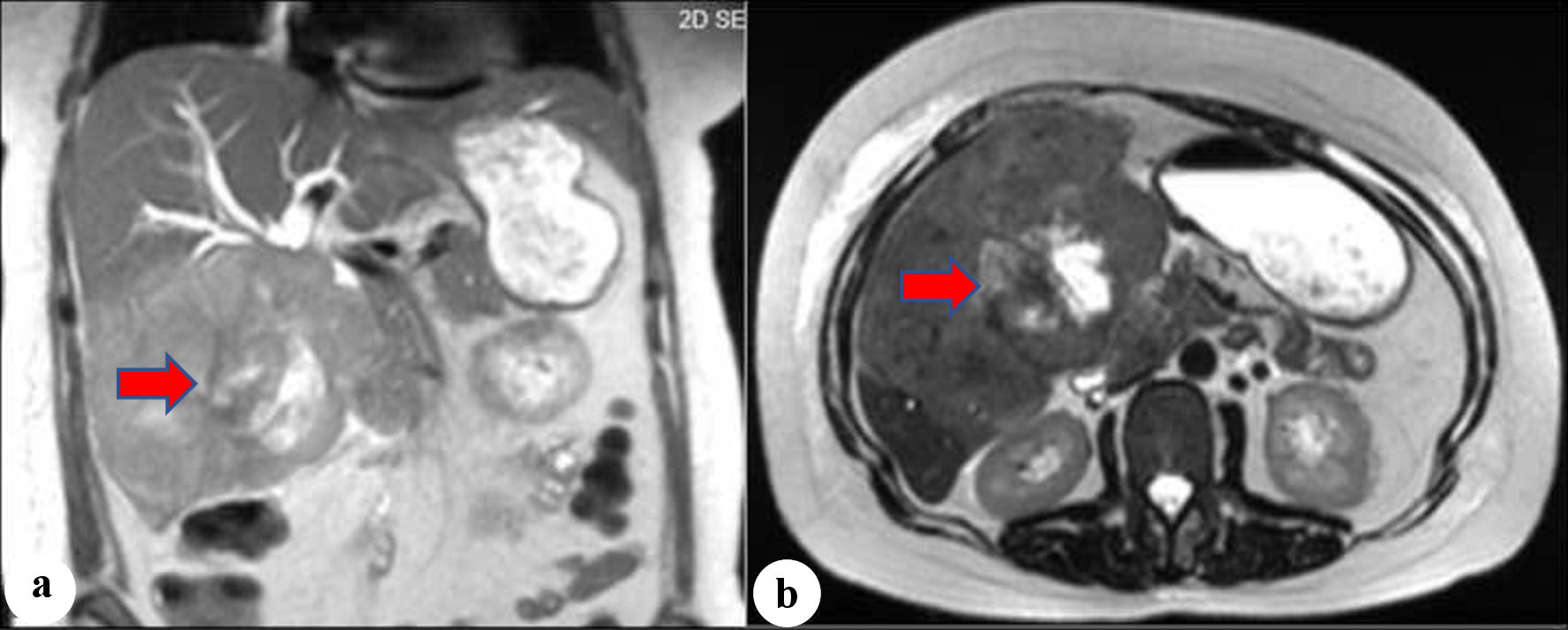
Figure 1. (a, b) Magnetic resonance imaging (MRI). The arrows show heterogeneous mass with a large necrotic center involving the right and left hepatic lobes, common hepatic duct, and proximal common bile duct.
Figure 2.
Figure 2. H&E staining images at × 20 showing squamous cell carcinoma, and the arrow shows focal keratinization. H&E: hematoxylin and eosin.
Figure 3.
Figure 3. H&E staining images (× 20) showing squamous cell carcinoma with necrosis. The arrow shows typical squamous cell carcinoma cells which are large with abundant eosinophilic cytoplasm with often vesicular, nuclei. No normal hepatic tissue is present in the biopsy. H&E: hematoxylin and eosin.
Figure 4.
Figure 4. Endoscopic retrograde cholangiopancreatography (ERCP). The arrow shows high-grade biliary stricture 3 - 4 cm involving the proximal common bile duct and common hepatic duct with significant intrahepatic dilation.