Gastroenterology Research, ISSN 1918-2805 print, 1918-2813 online, Open Access
Article copyright, the authors; Journal compilation copyright, Gastroenterol Res and Elmer Press Inc
Journal website https://www.gastrores.org

Case Report

Volume 15, Number 5, October 2022, pages 268-277


An Unusual Solitary Fibrous Tumor of the Ischiorectal Region

Figures

Figure 1.
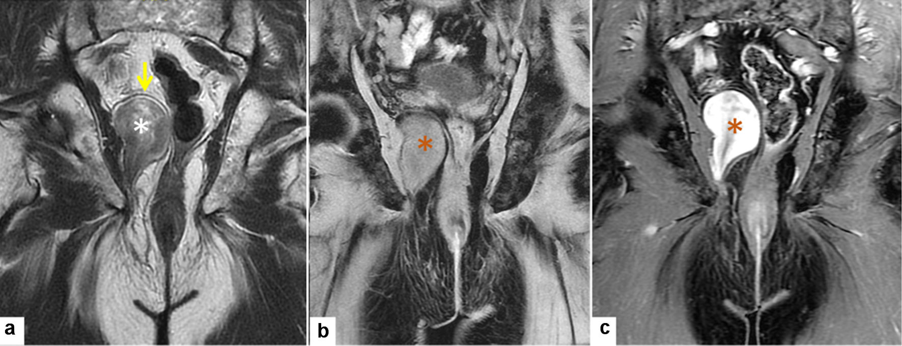
Figure 1. Radiological MRI features of solitary fibrous tumor (MRI: October 2018). (a) Coronal T2WI with fat saturation shows a well-defined heterogeneous predominantly low signal intensity right ischiorectal fossa ice-cone shaped mass (white asterisk). (b, c) Pre and post contrast-enhanced coronal T1WI with fat suppression showing homogenous lesion isointense to muscles in pre-contract and avidly enhancing following contrast administration (orange asterisk). MRI: magnetic resonance imaging.
Figure 2.
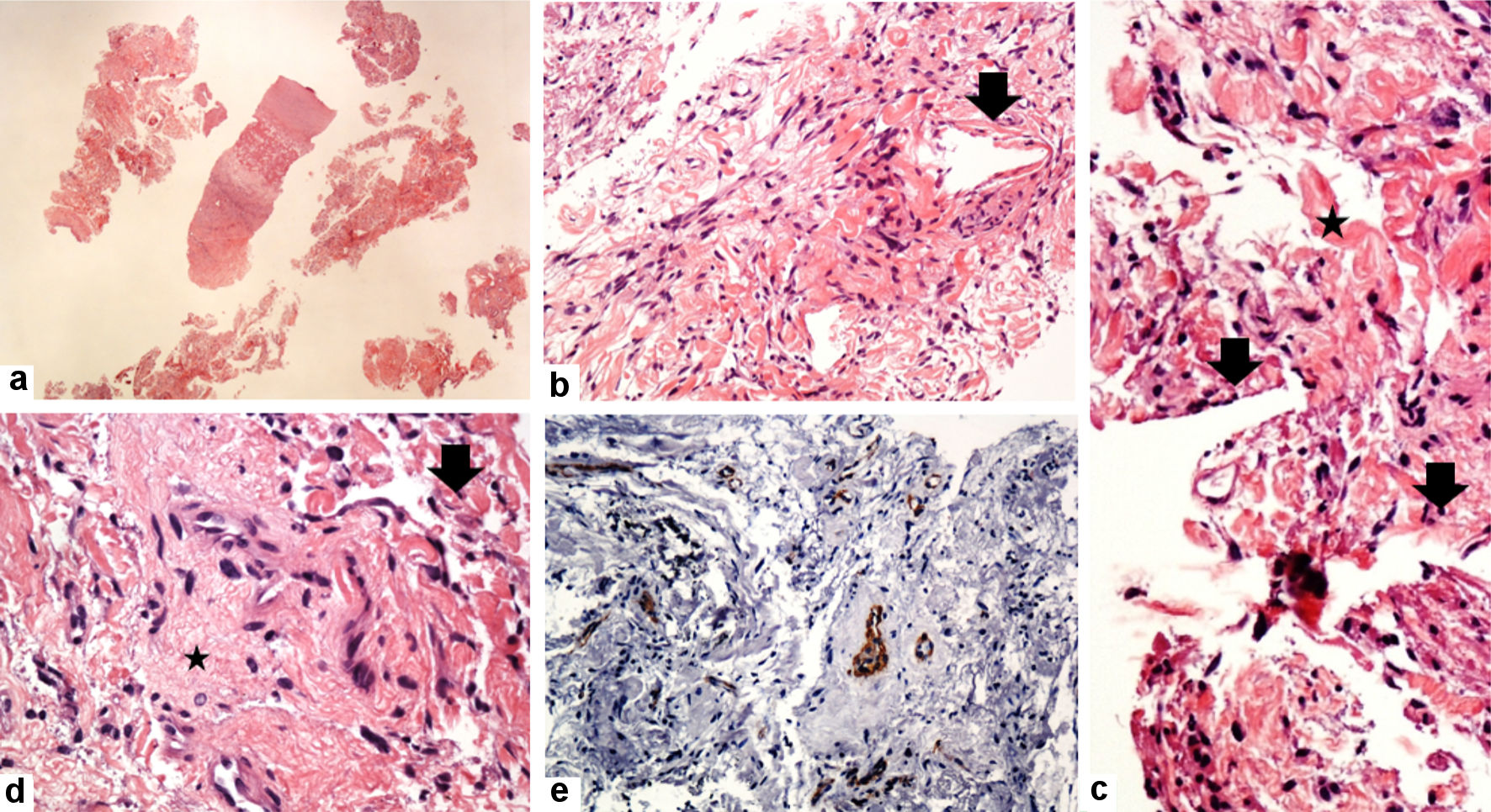
Figure 2. Histological features of solitary fibrous tumors (true cut needle biopsy: October 2018). (a-d) The tumor consists of a haphazard proliferation of short spindle to ovoid cells with banal-looking oval to spindle nuclei, pale eosinophilic cytoplasm, and some collagenized stroma (star), and the hemangiopericytoma-like vascular structures (arrow). The tumor cells are negative for SMA (e) (original magnifications: (a) × 20; (b) × 200; (c) × 400; (d) × 200; and (e) × 200).
Figure 3.
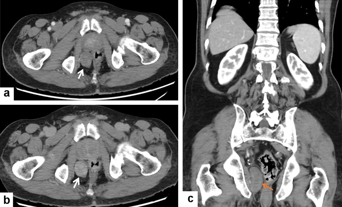
Figure 3. Radiological CT features of solitary fibrous tumor (CT: January 2022). (a, b) Contrast-enhanced CT of the pelvis shows a well-defined right ischiorectal fossa oval-shaped mass (white arrow) with progressive contrast enhancement on delayed CT (b). (c) Contrast-enhanced CT coronal reformat shows a well-defined elongated right ischiorectal fossa mass (orange arrow). CT: computed tomography.
Figure 4.
Figure 4. Radiological MRI features of solitary fibrous tumor (MRI: January 2022). (a) Coronal T2WI shows right well-defined heterogeneous predominantly low signal right ischiorectal fossa ice cone-shaped mass (white asterisk) upward displacing the right levator ani muscle (yellow arrow) without invasion. (b, c) Pre- and post-contrast-enhanced coronal T1WI with fat suppression show homogenous lesion isointense to muscles in pre-contract and avidly enhancing following contrast administration (orange asterisk). MRI: magnetic resonance imaging.
Figure 5.
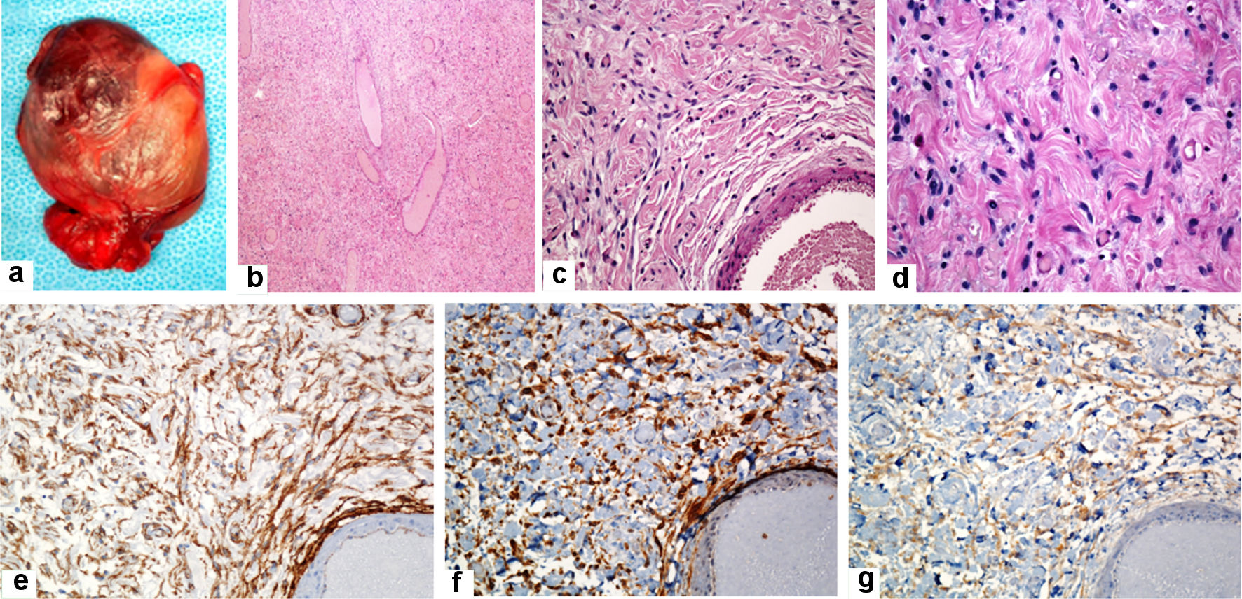
Figure 5. Gross and histological features of the solitary fibrous tumor (excisional biopsy: January 2022). Gross examination of the mass reveals well-defined, thinly encapsulated mass measuring about 5 × 3.5 × 3 cm (a). Histologic sections reveal cellular spindle cell neoplasm in a collagenous stroma with variable-sized blood vessels having stag-horn vascular morphology (b). The neoplastic cells are ovoid, short, or fusiform spindle-shaped cells with indistinct cell borders, bland looking nuclei, and indistinct nucleoli. They are arranged in short, poorly defined bundles, haphazardly fashion, and patternless pattern. The cells are streamed among the dermal collagens. No mitotic activity was seen. No significant nuclear atypia or necrosis was seen (c, d). The tumor cells are diffusely and strongly positive for CD34, BCL2, and CD99 (e, f, and g, respectively) and are negative for desmin, S100, and pancytokeratin (AE1/AE3) (original magnifications: (b) × 40, (c) × 200, (d) × 400, (e) × 200, (f) × 200, and (g) × 200).

Table

Table 1. Previous Studies About Pelvic Solitary Fibrous Tumor
 
StudiesAge/sexAnatomic siteClinical featuresTreatment
M: male; F: female; CT: computed tomography.
[14]Middle-agedIschiorectal fossaPelvic symptomatologyResection of the mass
[15]42/MIschiorectal fossaRectal massResection of the mass
[16]80/MThe perineal area close to the anal sphincterPerineal massResection of the mass
[19]62/MAnorectal region, recurrence in perineal regionAnorectal region, recurrence in perineal regionResection of the mass
[20]46/FPelvic mass displacing the rectumDoege-Potter syndromeResection of the mass
[21]27/FMesorectumIntraoperative pelvic tumorResection of the mass
[22]72/MRecurrent malignant tumor, ventral to sacral boneIntermittent loss of consciousnessResection of the mass
[23]64/MMass posterior to the bladder and associated with prostateLower abdominal painResection of the mass
[24]34/MMass close to the prostateCT findings suggestive of a prostatic mass lesionResection of the mass
[25]56/FMass in the mesorectumRadiological findings of giant pelvic massResection of the mass
[26]54/MWall of the low rectumColonoscopic studies suggestive of rectal massResection of the mass
[31]33/FTumor mass involving the sacral spinal canal and sacral foramenLower extremity pain, numbness, and muscle weaknessResection of the mass
[26]43/MLeft obturator areaAbdominal pain, appendicitisResection of the mass
[27]49/MMass related to the urinary bladderDifficulty in urinationResection of the mass
[28]76/MPelvic mass displacing the urinary bladder and rectumConstipation, abdominal pain, and urine retentionResection of the mass
[32]21/FMass arising from the serosa of the sigmoid colonAcute abdominal pain, constipation, and hematocheziaResection of the mass
[30]19/FMass in the ischioanal fossaPelvic pain and bleeding per rectumEmbolization and surgical resection