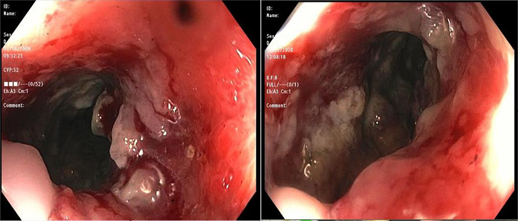
Figure 1. A colonoscopic image showing large circumferential ischemic, necrotic and friable areas in rectum.
| Gastroenterology Research, ISSN 1918-2805 print, 1918-2813 online, Open Access |
| Article copyright, the authors; Journal compilation copyright, Gastroenterol Res and Elmer Press Inc |
| Journal website https://www.gastrores.org |
Case Report
Volume 15, Number 2, April 2022, pages 106-111
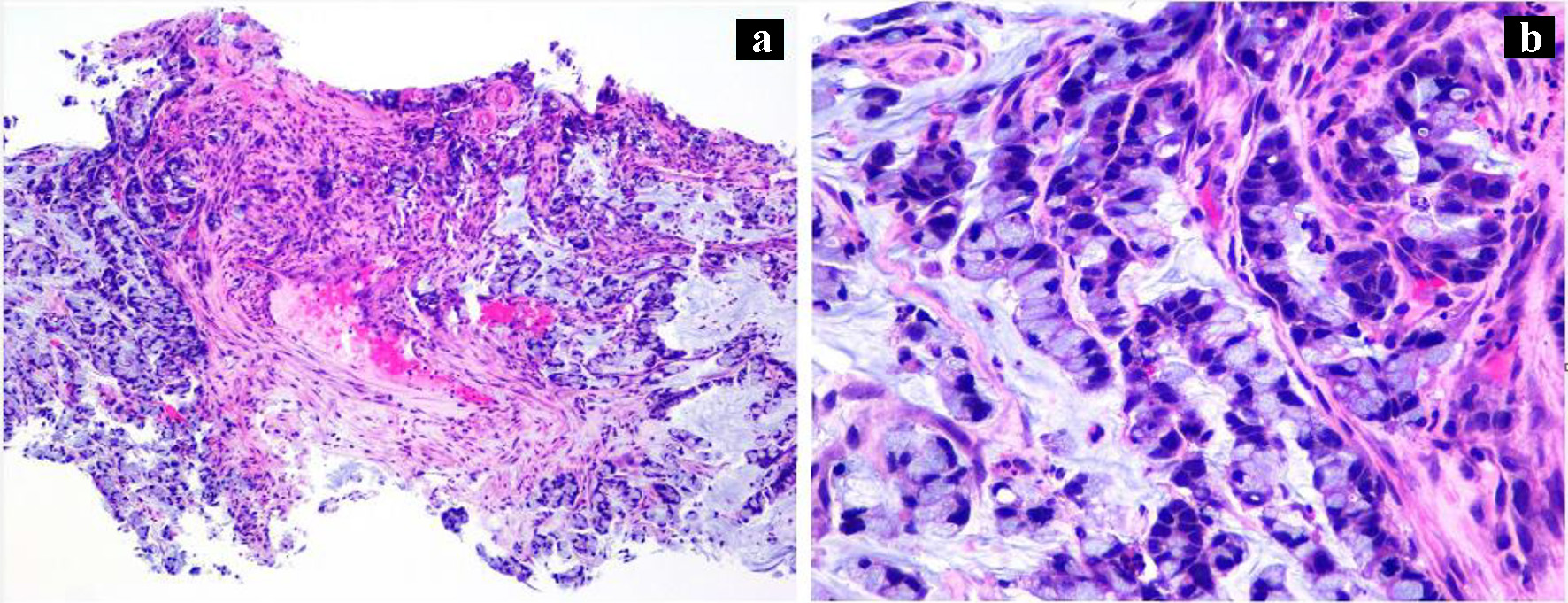
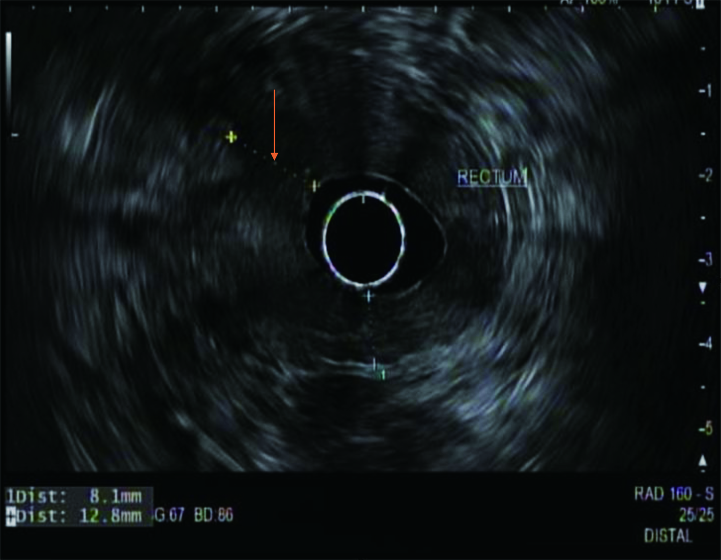
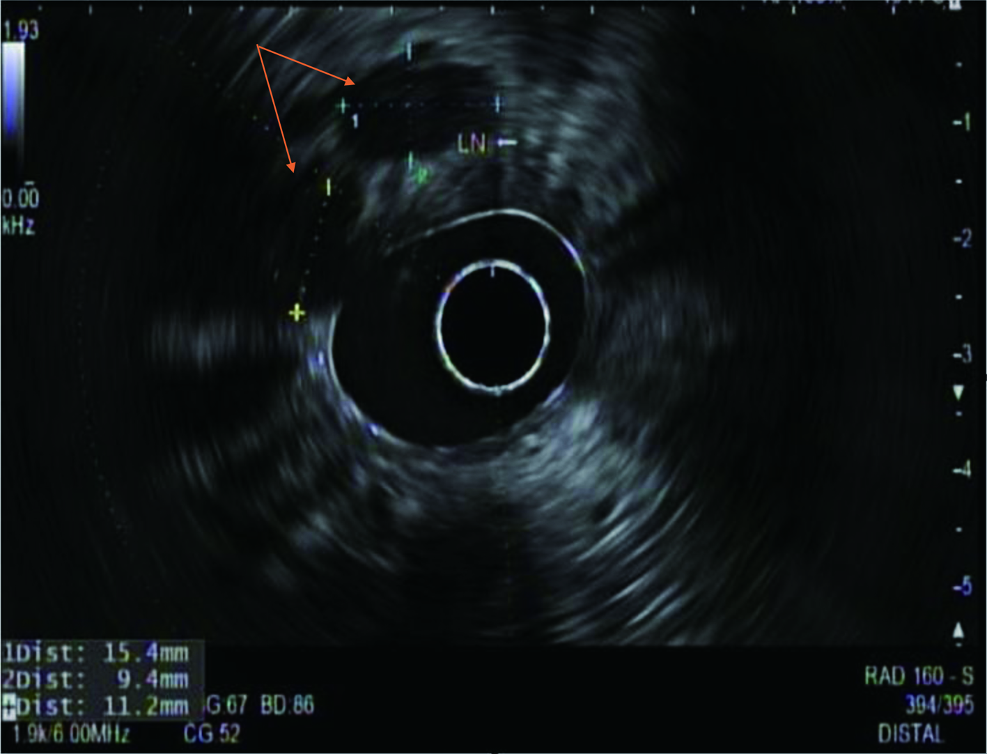
An Extremely Rare Case of Rectal Signet Ring Cell Carcinoma
Figures