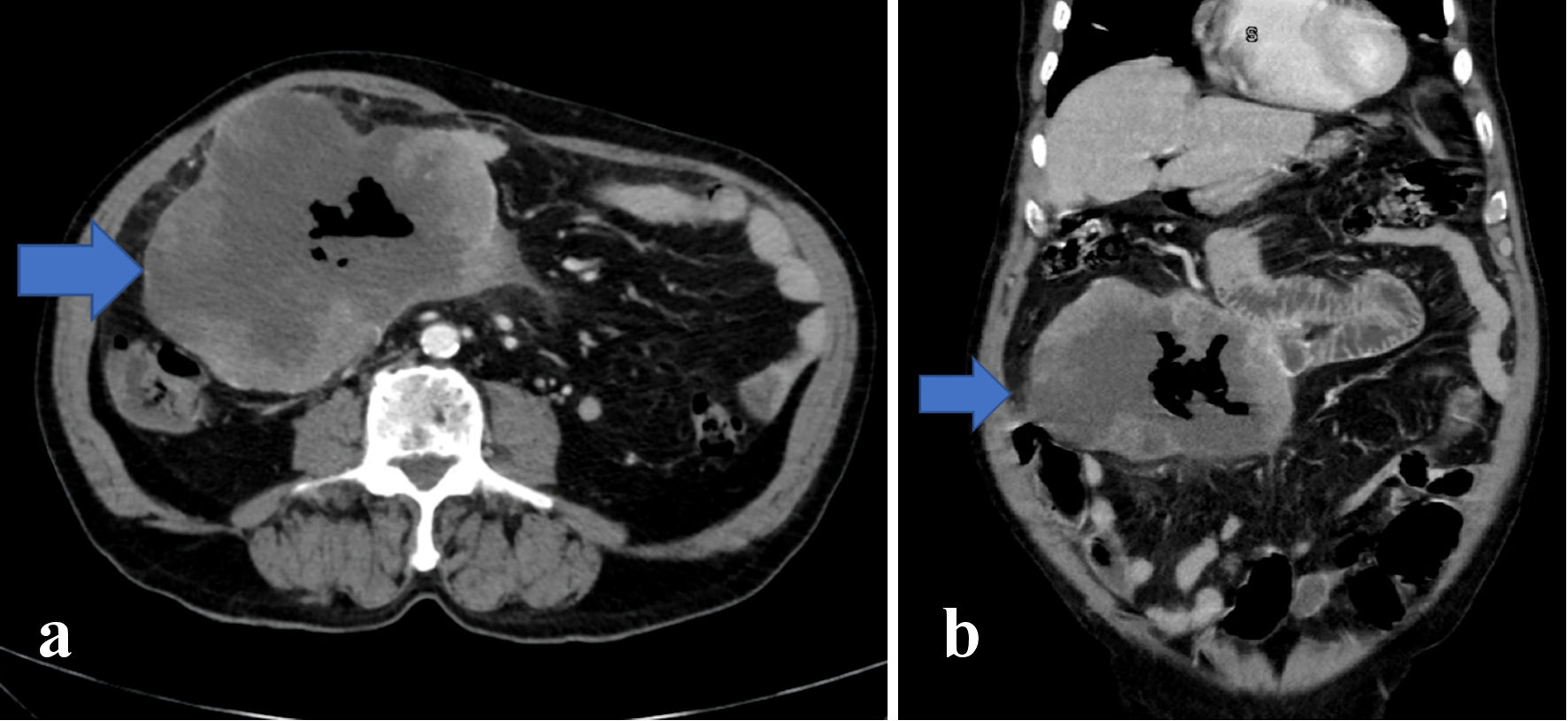
Figure 1. Axial (a) and coronal (b) images of abdominal CT scan indicate a sizeable mass (arrow) adjacent to the proximal duodenum with central necrosis and gas-containing, measuring approximately 11 × 14 × 10 cm. Also, distention of the bowels right adjacent to the mass is noticeable without proximal distension. CT: computed tomography.

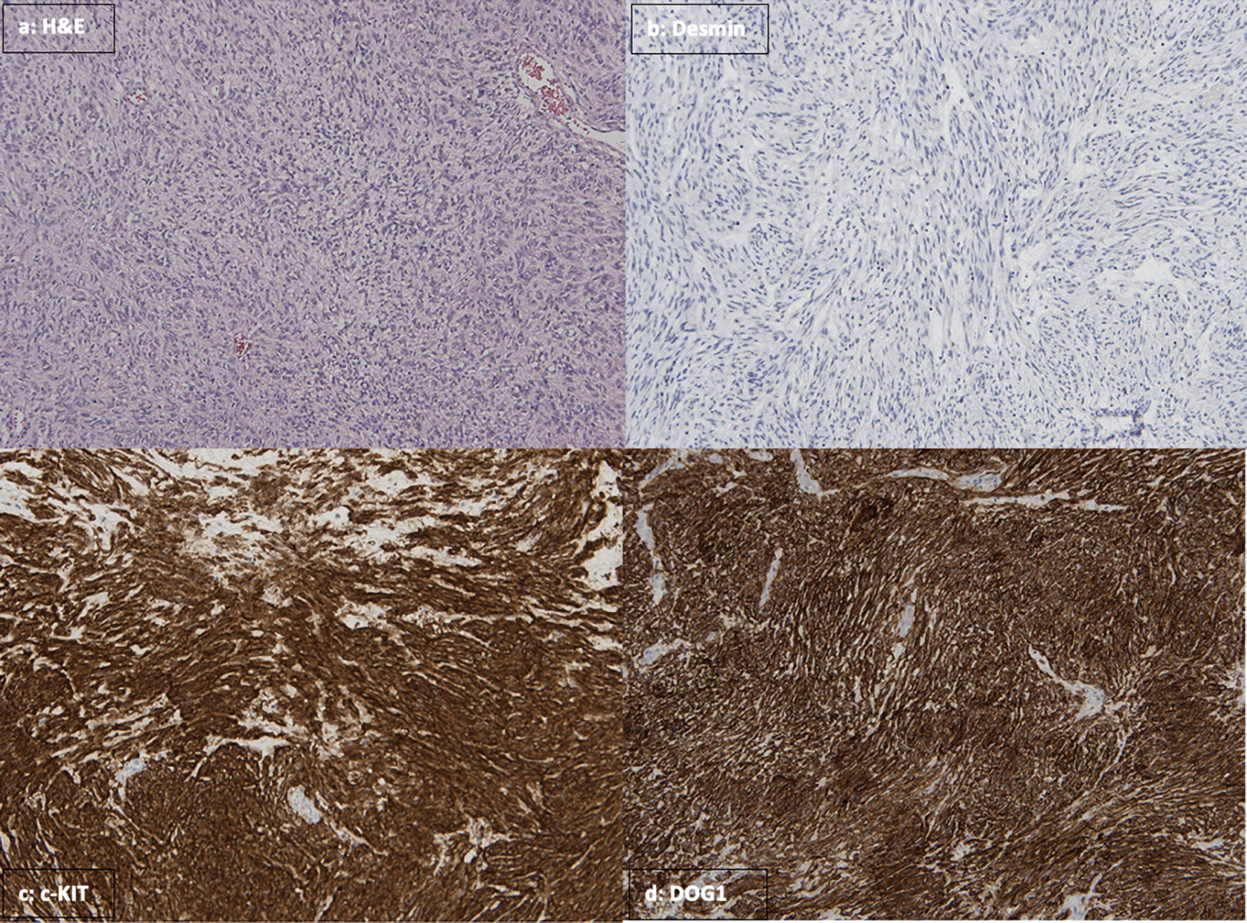
Figure 2. (a) In H&E stain, sections from the lesion showing the tumor spindle cell morphology and occasional mitoses. Immunohistochemical stains were performed, showing the tumor cells are negative for desmin (b), while strongly positive for c-kit/CD117 (c) and DOG1 (d). H&E: hematoxylin and eosin; DOG1: Discovered on GIST 1.

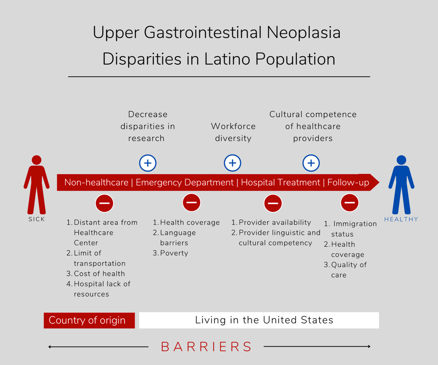
Figure 3. Barriers to access to health care encountered by the Latino population in their countries of origin and moving to the USA, including barriers to visit the emergency department, during hospitalization for treatment, and outpatient specialty clinic follow-up.


