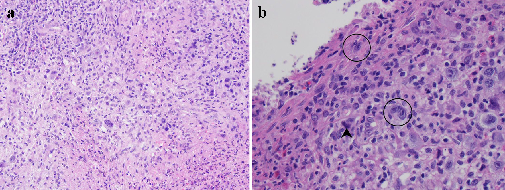
Figure 1. Histological evaluation of lesional tissue by hematoxylin and eosin (H&E) stain: (a) the lesion consists primarily of a destructive infiltrate of large, atypical cells mixed in a background of neutrophils, small lymphocytes, and eosinophils (× 100 magnification); (b) the large atypical cells resemble Hodgkin or Reed-Sternberg cells (circled) with 1 - 2 nuclei featuring vesicular chromatin and prominent inclusion-like nucleoli (× 400 magnification). Mummified cells are also depicted (arrowhead).
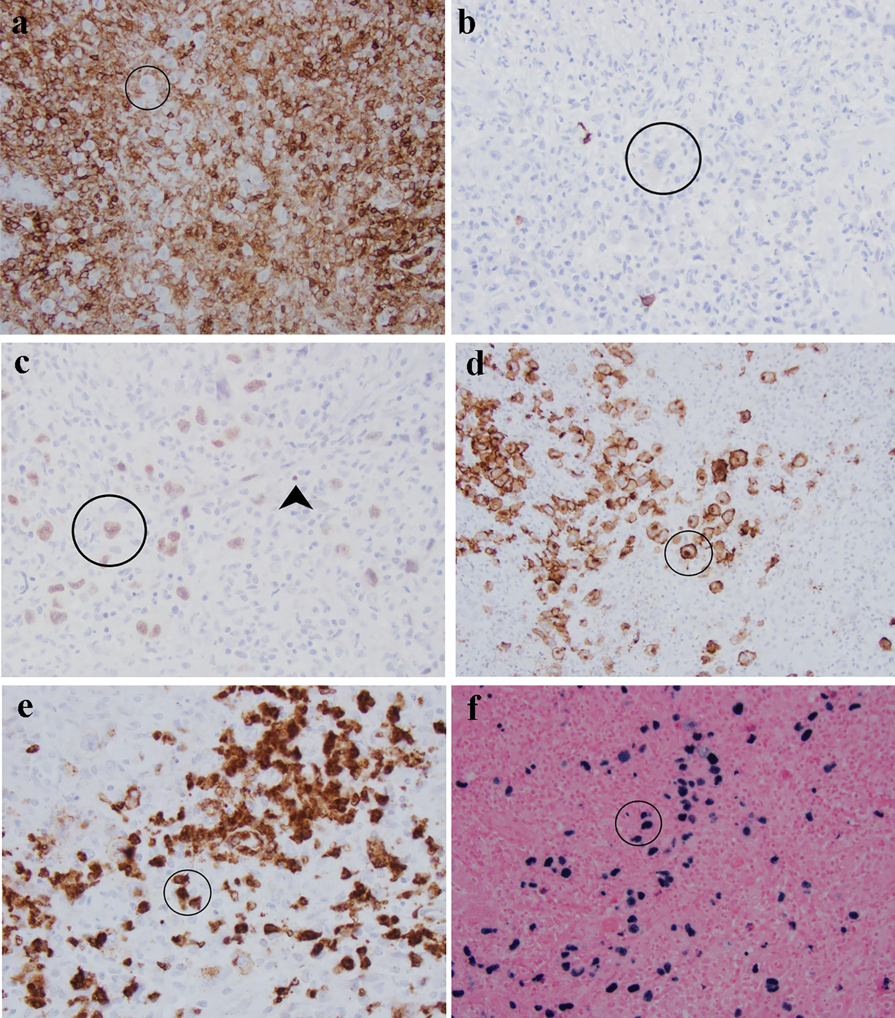
Figure 2. Characterization of atypical cells (circled) in this sigmoid mass by immunohistochemistry and in situ hybridization: (a) CD45 is negative in comparison with surrounding lymphocytes; (b) CD20; (c) Pax-5 is weak in Reed-Sternberg cells in comparison with strong staining in background B cells (arrowhead); (d) CD30 highlights membrane and Golgi bodies; (e) CD15 is positive in approximately 75% of Reed-Sternberg cells; (f) Epstein-Barr virus is positive in large cells rather than background small lymphocytes.