Gastroenterology Research, ISSN 1918-2805 print, 1918-2813 online, Open Access
Article copyright, the authors; Journal compilation copyright, Gastroenterol Res and Elmer Press Inc
Journal website https://www.gastrores.org

Review

Volume 14, Number 2, April 2021, pages 49-65


Idiopathic Non-Cirrhotic Portal Hypertension and Porto-Sinusoidal Vascular Disease: Review of Current Data

Figures

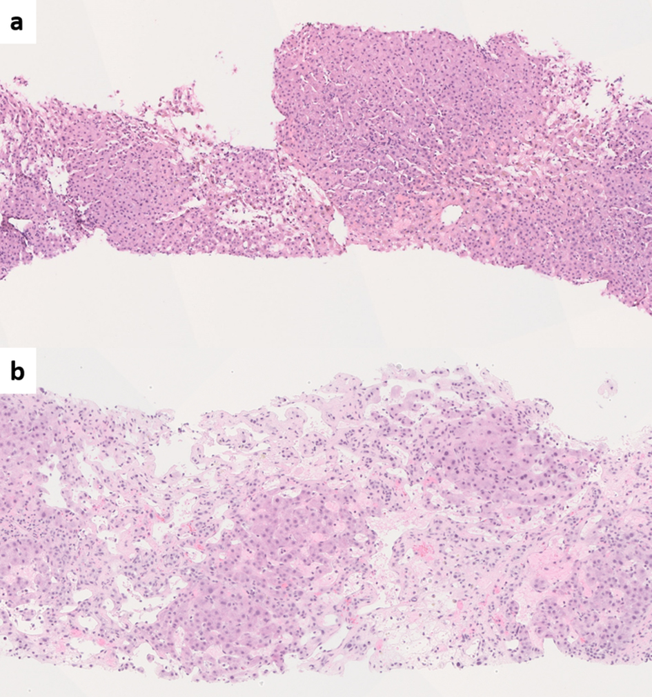
Figure 1.
Figure 1. (a) Liver biopsy from Budd-Chiari syndrome (BCS) shows venous outflow obstruction pattern injury with mild centrizonal sinusoidal dilatation and congestion (hematoxylin and eosin (H&E), × 100). (b) Chronic BCS with extensive sinusoidal dilatation, centrizonal hepatocyte atrophy and dropout and fibrosis. The portal tracts are relatively spared (H&E, × 100).
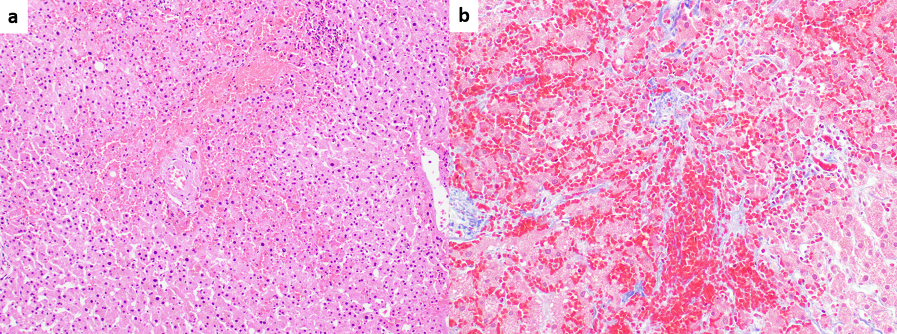
Figure 2.
Figure 2. (a) Sinusoidal obstruction syndrome (SOS) associated with oxaliplatin chemotherapy. Marked centrizonal congestion and sinusoidal widening is noted (hematoxylin and eosin, × 100). (b) Trichrome stain shows perisinusoidal fibrosis (Masson-trichrome, × 200).
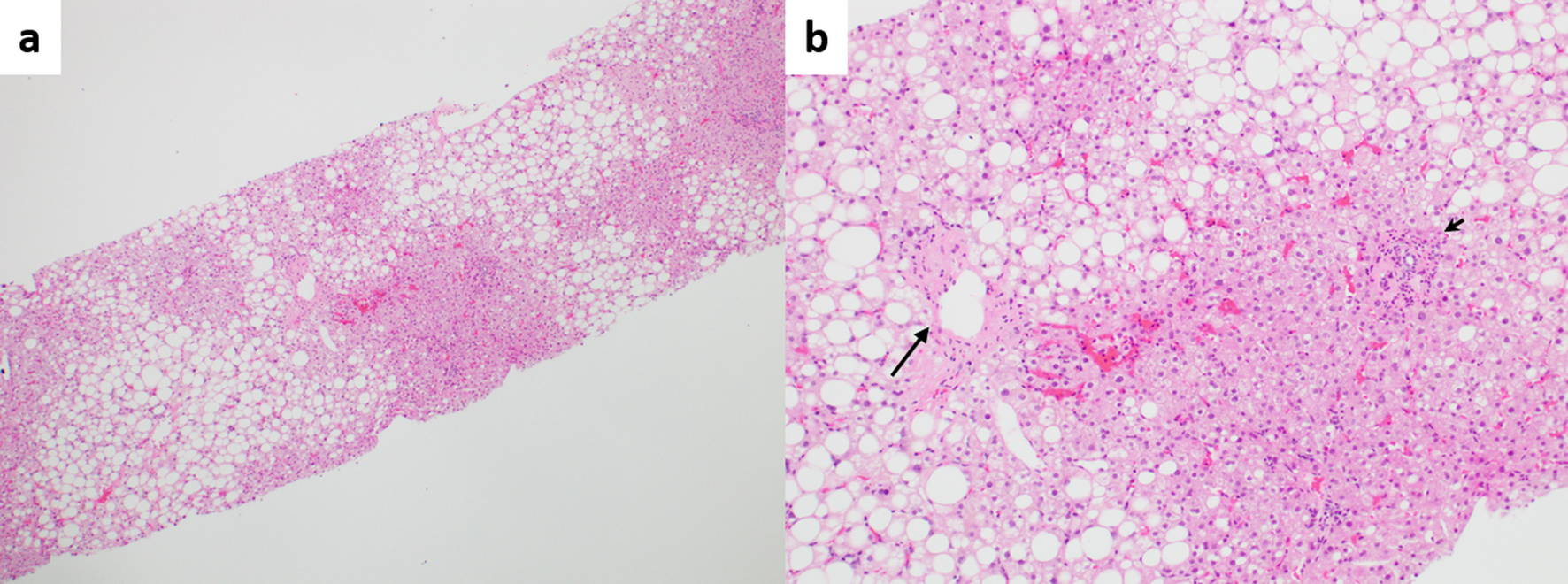
Figure 3.
Figure 3. (a) Fatty liver disease without cirrhosis or advanced fibrosis, but with portal hypertension. Zonal steatosis is noted (hematoxylin and eosin (H&E), × 40). (b) Higher magnification view showing pericentral fibrosis (long arrow) probably secondary to fatty liver disease, and phlebosclerosis (obliterative portal venopathy, short arrow), possibly secondary to concurrent porto-sinusoidal vascular disease (H&E, × 100).
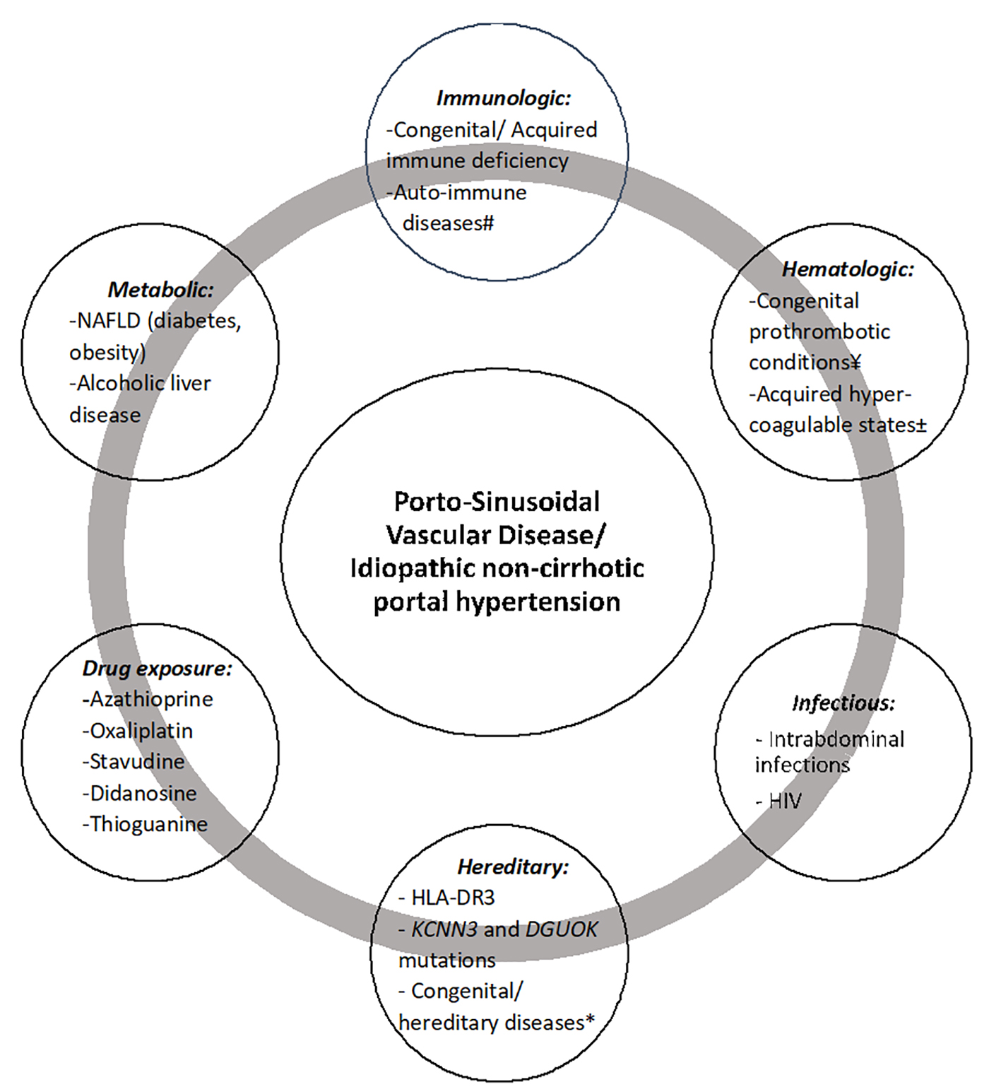
Figure 4.
Figure 4. Etiopathogenic associations with porto-sinusoidal vascular disease. #Systemic lupus erythematosus, rheumatoid arthritis, scleroderma, Sjogren’s syndrome, inflammatory bowel diseases, celiac disease, autoimmune hepatitis, Felty’s syndrome. ¥Protein C or S deficiency, factor V Leiden, factor II mutation, antithrombin III deficiency. ±Antiphospholipid syndrome, malignancy, ADAMTS 13 deficiency, oral contraceptive use. *Turner syndrome, Adams-Oliver syndrome, cystic fibrosis, phosphomannose isomerase deficiency. NAFLD: non-alcoholic fatty liver disease; HIV: human immunodeficiency virus.
Figure 5.
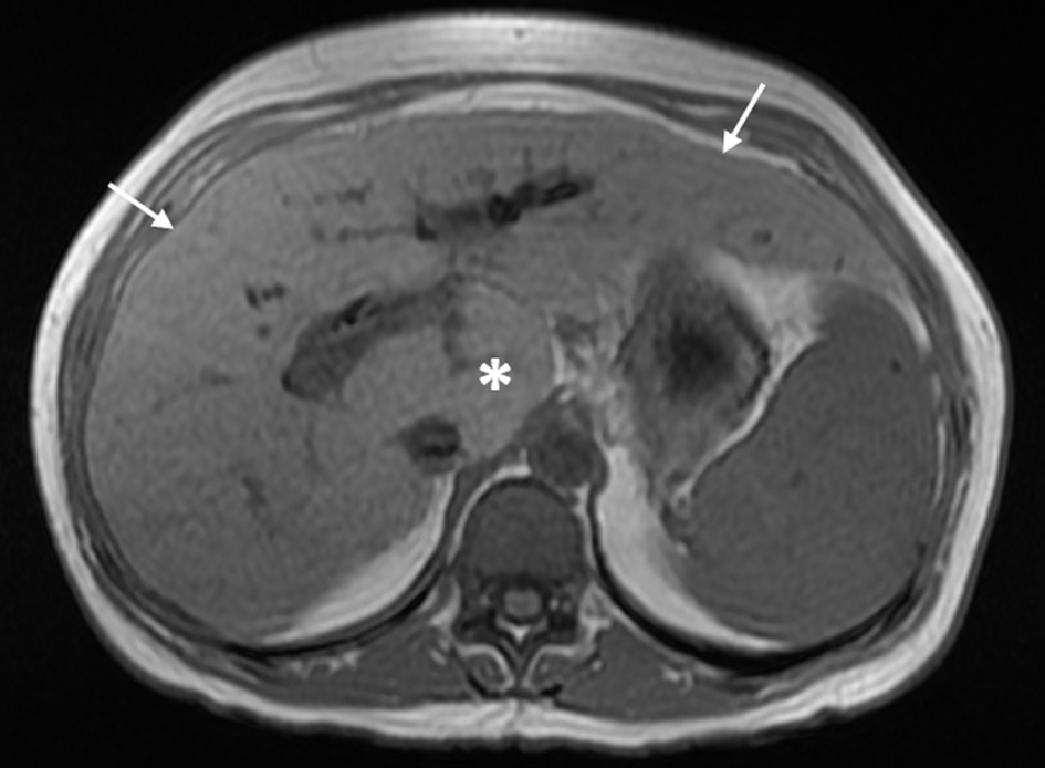
Figure 5. Magnetic resonance imaging with contrast shows mild nodular contour of the liver surface (arrows) and relative hypertrophy of the caudate lobe (*) in porto-sinusoidal vascular disease.
Figure 6.
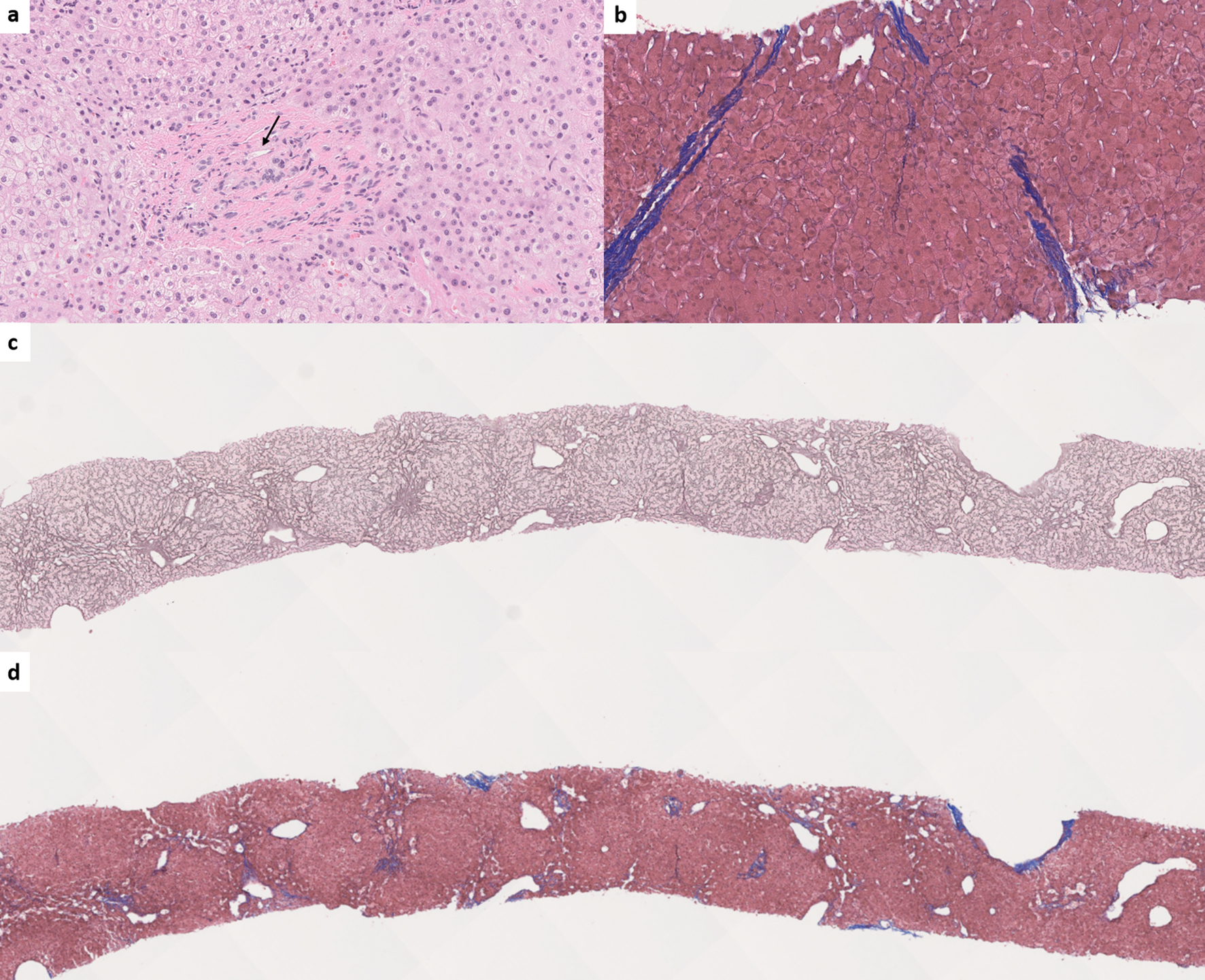
Figure 6. (a) Obliterative portal venopathy (also known as phlebosclerosis) with inconspicuous portal venous branch (arrow) (hematoxylin and eosin, × 200). (b) Incomplete septal fibrosis or cirrhosis (Masson-trichrome, × 150). (c) Nodular regenerative hyperplasia. The nodularity is highlighted by reticulin special stain (reticulin, × 30). (d) Trichrome stain shows the absence of cirrhosis in nodular regenerative hyperplasia (Masson-trichrome, × 30).
Figure 7.
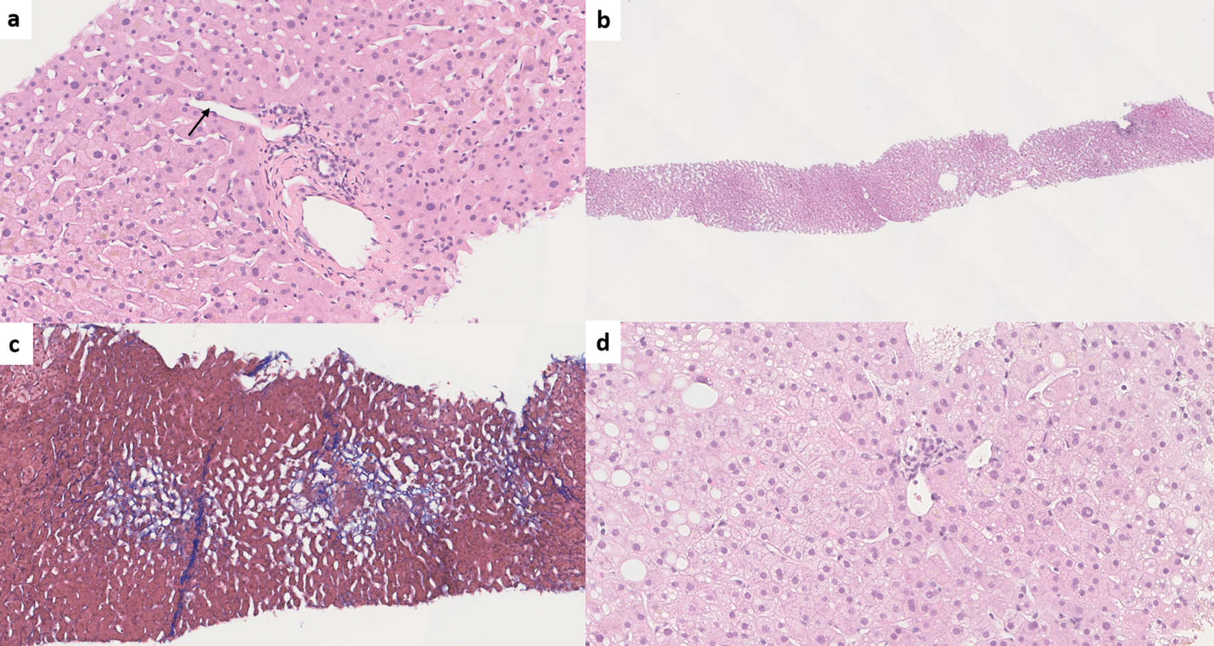
Figure 7. (a) Paraportal shunt vessel (arrow) in idiopathic non-cirrhotic portal hypertension/porto-sinusoidal vascular disease (INCPH/PSVD) (hematoxylin and eosin (H&E), × 200). (b) Irregularly distributed portal tracts and central veins in INCPH/PSVD. Non-zonal sinusoidal dilatation is also noted (H&E, × 50). (c) Mild perisinusoidal fibrosis (Masson-trichrome, × 130). (d) Rudimentary portal tract in INCPH/PSVD (H&E, × 250).
Figure 8.
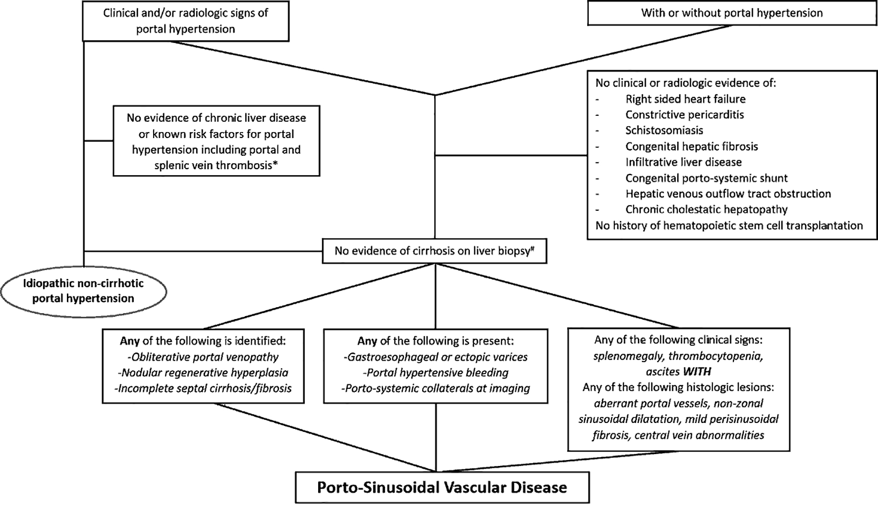
Figure 8. Diagram summarizing the diagnostic approach for idiopathic non-cirrhotic portal hypertension and porto-sinusoidal vascular disease. *Portal vein thrombosis may be seen along the natural disease course of idiopathic non-cirrhotic portal hypertension. #Biopsy adequacy criteria are available for porto-sinusoidal vascular disease (core liver biopsy ≥ 20 mm in length or featuring ≥ 10 portal tracts, or when considered adequate by an expert pathologist).

Tables

Table 1. Symptoms and Signs of Portal Hypertension
 
Specific signsNon-specific signs
HPVG: hepatic venous pressure gradient.
ClinicalVariceal bleeding, collaterals seen on physical examinationAscites, thrombocytopenia, splenomegaly
EndoscopicVarices
RadiographicPorto-systemic collaterals, increased HVPGAscites, splenomegaly


Table 2. Histologic Features of Idiopathic Non-Cirrhotic Portal Hypertension/Porto-Sinusoidal Vascular Disease and Their Definitions [1, 2, 8-11]
 
Specific features*
*Specific and not specific features (lesions) are determined by Vascular Liver Disease Interest Group. This categorization is applicable to porto-sinusoidal vascular disease only [2]. Specificity of these histologic lesions is unknown and not defined for idiopathic non-cirrhotic portal hypertension [11].
  Obliterative portal venopathyWall thickening and fibrosis with luminal narrowing, obliteration and eventual loss of intrahepatic portal vein branches
  Nodular regenerative hyperplasiaMicronodularity of hepatic parenchyma in the absence of liver fibrosis. Nodules are composed of hyperplastic central zones and peripheral atrophic hepatic cell plates.
  Incomplete septal fibrosis/cirrhosisDelicate fibrous septa originating from a portal tract and ending blindly within a hepatic lobule without clear connection to central veins or other portal tracts
Not specific features*
  Lobular changes
    Sinusoidal dilatationSinusoidal lumen wider than one liver cell plate in the absence of artifactual tearing, usually non-zonal
    MegasinusoidsSevere sinusoidal dilatation with cystic blood lake formation. Some authors used the term to describe dilated periportal shunting vessels.
    Perisinusoidal fibrosisStellate pattern of collagen deposition around hepatic sinusoids highlighted by collagen stain
    Central vein abnormalitiesCentral vein dilatation, pericentral vein fibrosis, multiplicity of central veins per lobule
  Portal tract changes
    Periportal shunting vesselsSingle or multiple thin-walled vascular channels seen outside but in contact with a portal tract
    Herniated portal veinPortal vein branch, often dilated, abutting the adjacent hepatic parenchyma at limiting plate without a rim of intervening connective tissue
    Portal tract remnantPortal tract smaller than twice the diameter of a bile duct, often with an inconspicuous/absent portal vein branch or herniated portal vein
    Increased arteriole profilesArterialized portal venous branches with acquired smooth muscle layer
    Multiplicity of portal veinsIncreased number of portal vein branches within a portal tract, also known as angiomatous transformation


Table 3. Different Terminologies Used for Idiopathic Non-Cirrhotic Portal Hypertension/Porto-Sinusoidal Vascular Disease
 
TerminologiesReferences
Idiopathic portal hypertensionKobayashi et al (1976) [19], Okuda et al (1982) [22], Nakanuma et al (1989) [23], Saito et al (1993) [24], Oikawa et al (1998) [25], Yamaguchi et al (1999) [26], Okudaira et al (2002) [27], Tsuneyama et al (2002) [28], Kogawa et al (2005) [29], Matsutani et al (2005) [30], Chang et al (2009) [31], Seijo et al (2012) [32], Furuichi et al (2013) [33], Siramolpiwat et al (2014) [34], Kotani et al (2015) [35]
Non-cirrhotic portal fibrosisSarin et al (1987) [36], Mukta et al (2017) [20], Sood et al (2017) [37]
Obliterative portal venopathyMikkelsen et al (1965) [38], Nayak et al (1969) [39], Cazals-Hatem et al (2011) [40], Glatard et al (2012) [41], Aggarwal et al (2013) [42], Franchi-Abella et al (2014) [43], Arora et al (2015) [44], Guido et al (2016) [12], Besmond et al (2018) [45]
Hepatoportal sclerosisMikkelsen et al (1965) [38], Girard et al (2005) [46], Fiel et al (2007) [47], Krishnan et al (2012) [48]
Benign intrahepatic portal hypertensionLevison et al (1982) [49]
Intrahepatic non-cirrhotic portal hypertensionKingham et al (1981) [50], Krasinskas et al (2005) [6], Eapen et al (2011) [51]
Non-cirrhotic portal hypertensionOhbu et al (1994) [52], Nakanuma et al (1996) [16], Rajekar et al (2011) [3], Rajesh et al (2018) [53], Gioia et al (2018) [54], Nicoara-Farcau et al (2020) [55], Gioia et al (2020) [56]
Idiopathic non-cirrhotic intrahepatic portal hypertensionHillaire et al (2002) [21], Goel et al (2011) [57]
Partial nodular transformationSherlock et al (1966) [58], Wanless et al (1985) [59]
Nodular regenerative hyperplasiaSteinert et al (1959) [60], Wanless et al (1990) [61], Radomski et al (2000) [62], Austin et al (2004) [63], Malamut et al (2008) [64], Leung et al (2009) [65], Jharap et al (2015) [66]
Idiopathic presinusoidal portal hypertensionPolish et al (1962) [67]
Incomplete septal cirrhosisSciot et al (1988) [68]