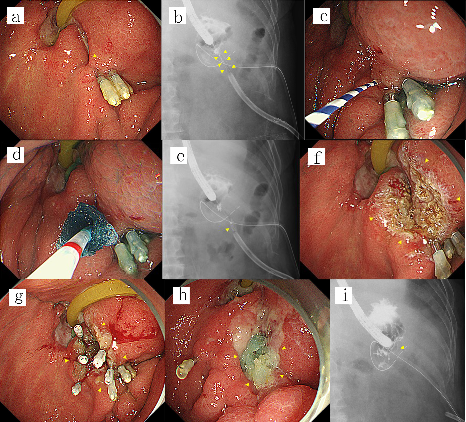
Figure 1. (a) A fistula was endoscopically detected at a gastrojejunal anastomosis (arrowhead) after failed fistula closure using hemoclips. (b, c) Contrast imaging performed by introducing contrast medium through the orifice of the fistula revealed an anastomo-cutaneous fistula (arrowheads). A guidewire was then introduced into the fistula (arrowhead). (d, e) A small piece of PGA sheet was skewered onto the guidewire at the center and then pushed using the tapered catheter over the guidewire and delivered into the fistula (arrowhead). This procedure was repeated five times while adjusting the depth of the PGA sheets on a radiogram. (f) The orifice of the fistula along with the surrounding mucosa was ablated using argon plasma coagulation (arrowheads). (g) The orifice of the fistula along with the surrounding mucosa was shielded by a piece of PGA sheet fixed with five hemoclips and fibrin glue (arrowheads). (h, i) The fistula was not detectable either endoscopically or on contrast radiograms by adding one more application 7 days after the first procedure (arrowheads), and the fistula remained closed until the patient died 465 days after the last procedure. PGA: polyglycolic acid.