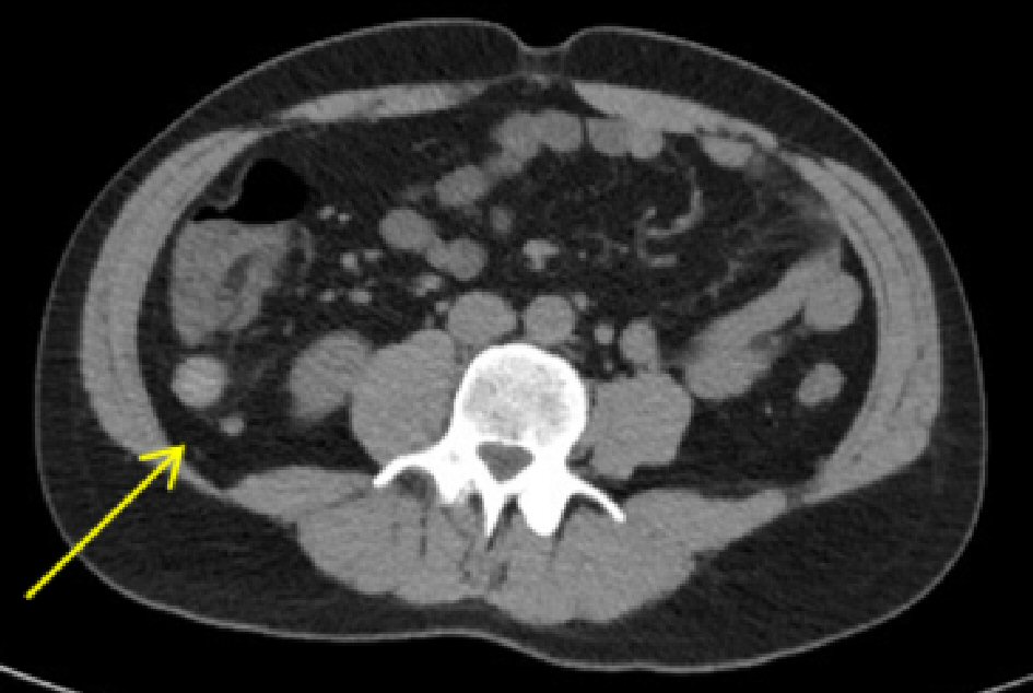
Figure 1. Abdominal CT imaging demonstrated a 3.7-cm mass at the appendiceal tip (arrow). CT: computed tomography.
| Gastroenterology Research, ISSN 1918-2805 print, 1918-2813 online, Open Access |
| Article copyright, the authors; Journal compilation copyright, Gastroenterol Res and Elmer Press Inc |
| Journal website http://www.gastrores.org |
Case Report
Volume 13, Number 2, April 2020, pages 85-87
First Reported Case of Extramedullary Plasmacytoma of the Appendix
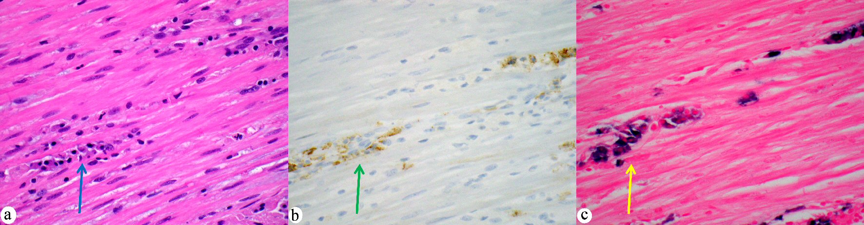
Figures