Gastroenterology Research, ISSN 1918-2805 print, 1918-2813 online, Open Access
Article copyright, the authors; Journal compilation copyright, Gastroenterol Res and Elmer Press Inc
Journal website http://www.gastrores.org

Case Report

Volume 12, Number 6, December 2019, pages 315-319


Spontaneous Perforation of an Acalculous Hydropic Gallbladder in a Diabetic Patient With Neuropathy: An Underdiagnosed Entity

Figures

Figure 1.
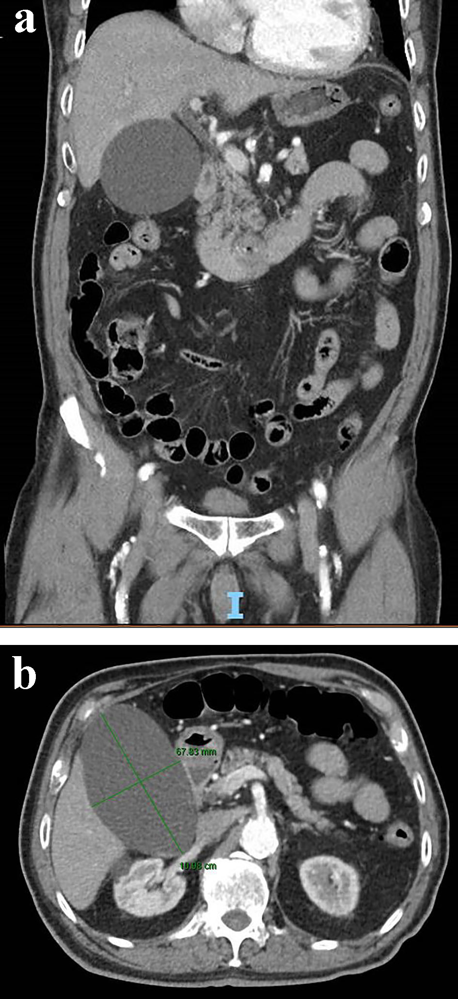
Figure 1. Coronal (a) and axial view (b) of a contrast-enhanced abdominal CT scan showing an acalculous hydropic gallbladder. CT: computed tomography.
Figure 2.
Figure 2. Ultrasound imaging of an acalculous hydropic gallbladder with demonstration of gallbladder sludge and without evidence of gallbladder wall thickening or pericholecystic fluid.
Figure 3.
Figure 3. Hepatobiliary iminodiacetic acid (HIDA) scan with visualization of the gallbladder within 15 min post-injection of bright nuclear material. There is also early evidence of biliary leak into the right peritoneal cavity at 45 min which increases up to 4 h post-contrast admission, which suggests a perforated gallbladder.
Figure 4.
Figure 4. Axial view of a contrast-enhanced CT of the abdomen demonstrating a decompressed gallbladder and biliary leakage. CT: computed tomography.