Figures
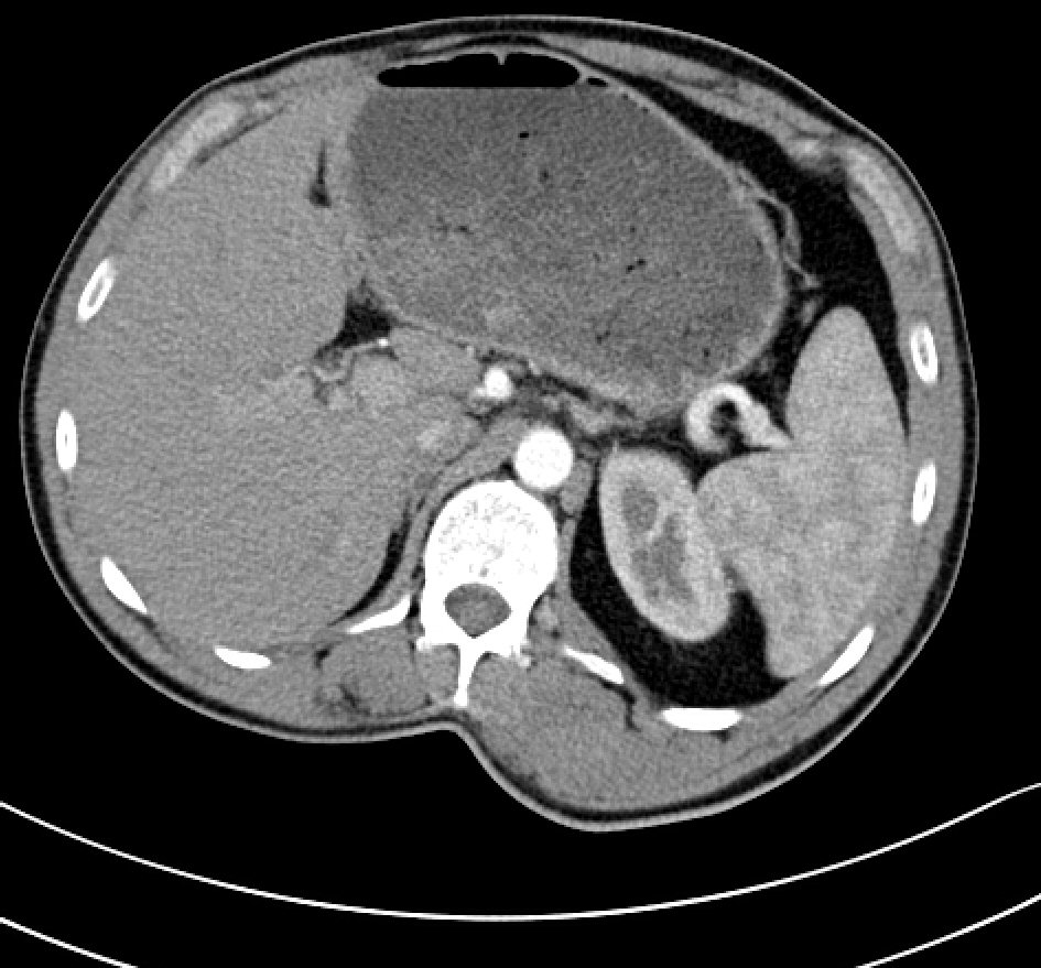
Figure 1. CT scan demonstrates gastrostasis.
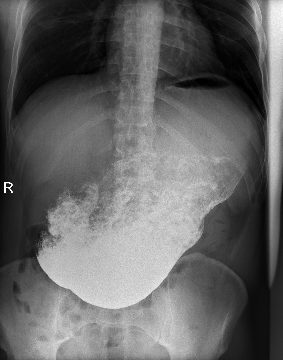
Figure 2. X-ray examination: barium sulphate was used as contrast agent. Repeated examination 30 min after barium administration demonstrates gastrostasis.
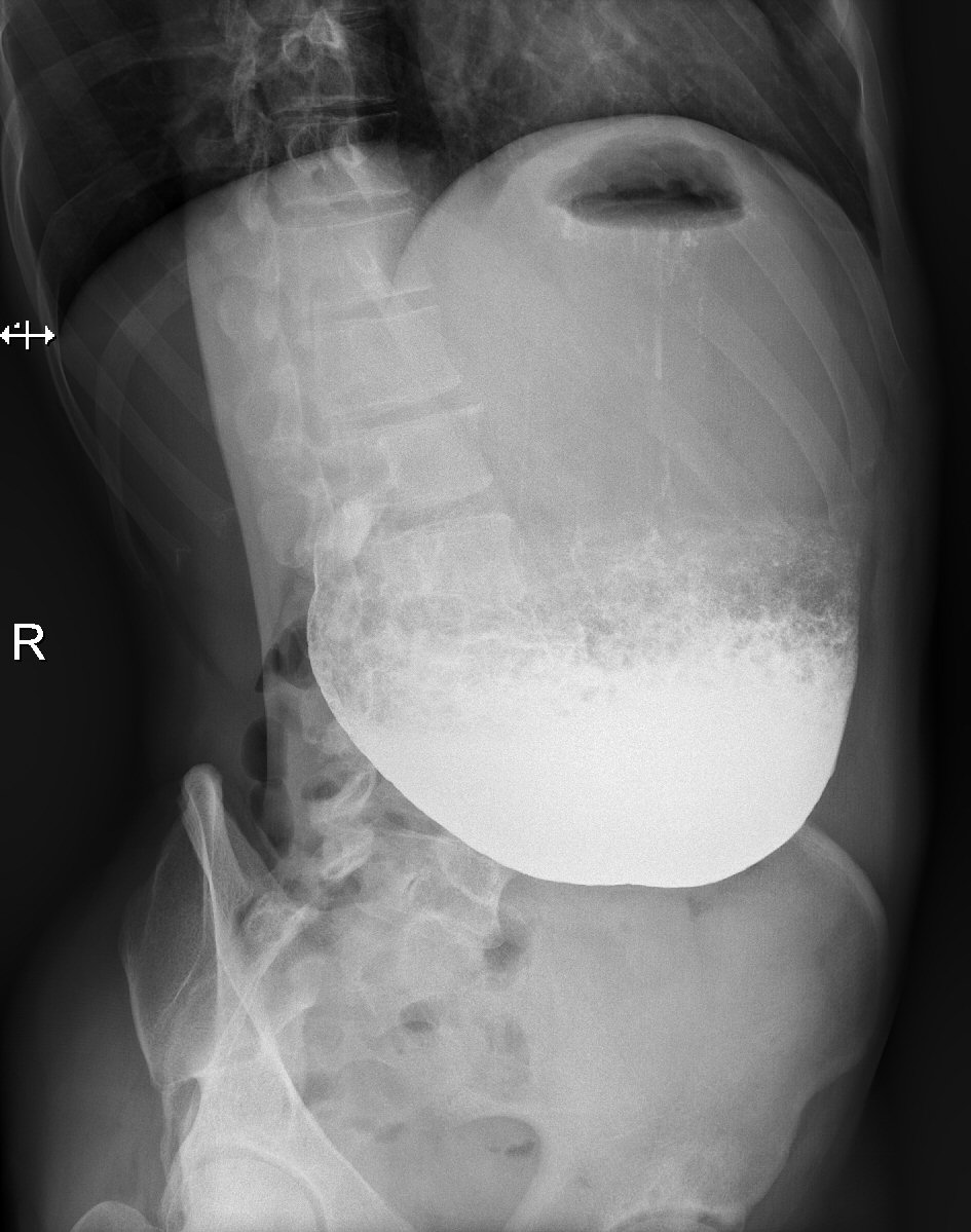
Figure 3. X-ray examination: 30 min after barium administration, lateral oblique projection.
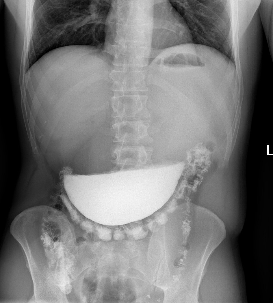
Figure 4. X-ray examination demonstrates stasis of the contrast agent, minimal amount of contrast agent passed through the pylorus.
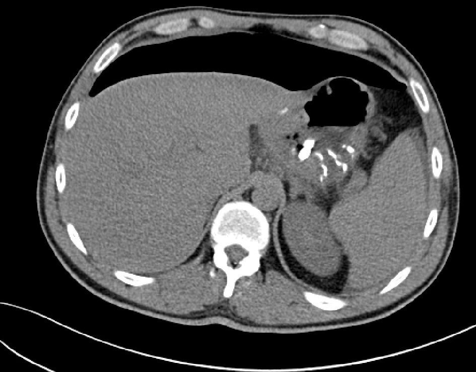
Figure 5. CT examination after partial resection of the stomach, the examination demonstrated only minimal amount of contrast agent intraperitoneally outside the gastrointestinal tract; and an active leak was not identified.
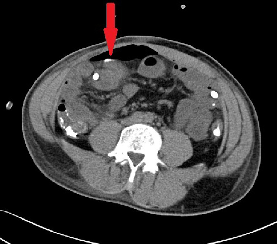
Figure 6. CT examination after partial resection of the stomach, small amount of contrast agent intraperitoneally outside the digestive tract is marked by the arrow.