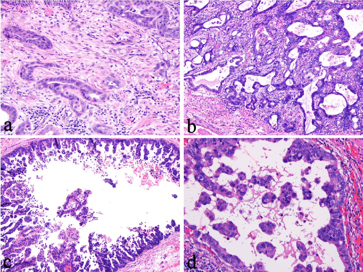
Figure 1. (a) Desmoplasia in an esophageal intramucosal adenocarcinoma (IMAC) (400 ×). (b) Myxoid stromal changes in an esophageal IMAC (100 ×). (c, d) Detached papillae in an esophageal IMAC (100 × and 200 ×).
| Gastroenterology Research, ISSN 1918-2805 print, 1918-2813 online, Open Access |
| Article copyright, the authors; Journal compilation copyright, Gastroenterol Res and Elmer Press Inc |
| Journal website http://www.gastrores.org |
Original Article
Volume 12, Number 2, April 2019, pages 72-77
Desmoplasia and Detached Papillae in Early Esophageal Adenocarcinoma: A Histologic Study on Endoscopic Submucosal Dissection Specimens
Figures
Tables
| LVI N (%) | P value | |
|---|---|---|
| IMAC | ||
| Desmoplasia (n = 5) | 4 (80) | 0.048 |
| Desmoplasia-negative (n = 16) | 4 (25) | |
| SMAC | 0.24 | |
| Desmoplasia (n = 11) | 8 (73) | |
| Desmoplasia-negative (n = 4) | 1 (25) | |
| IMAC and SMAC combined | 0.006 | |
| Desmoplasia (n = 16) | 12 (75) | |
| Desmoplasia-negative (n = 20) | 5 (25) |
| LVI N (%) | P value | |
|---|---|---|
| IMAC | ||
| Presence of detached papillae (n = 15) | 7 (46.7) | 0.34 |
| Absence of detached papillae (n = 6) | 1 (16.7) | |
| SMAC | 0.24 | |
| Presence of detached papillae (n = 11) | 8 (72.3) | |
| Absence of detached papillae (n = 4) | 1 (25) | |
| IMAC and SMAC combined | 0.06 | |
| Presence of detached papillae (n = 26) | 15 (57.7) | |
| Absence of detached papillae (n = 10) | 2 (20) |
| IMAC (OR: 95% CI) | SMAC (OR: 95% CI) | IMAC and SMAC (OR: 95% CI) | |
|---|---|---|---|
| OR: odds ratio; NA: not applicable. | |||
| Age | 1.09 (0.98 - 1.2), P = 0.1 | 0.88 (0.73 - 1.1), P = 0.2 | 1.03 (0.96 - 1.1), P = 0.4 |
| Gender (F/M) | NA | NA | 0.53 (0.04 - 6.4), P = 0.6 |
| Largest dimension of ESD specimen | 0.5 (0.23 - 1.1), P = 0.09 | 1.07 (0.58 - 2), P = 0.8 | 0.73 (0.47 - 1.1), P = 0.2 |
| Average thickness of resection | 1.29 (0.64 - 2.6), P = 0.5 | NA | 1.27 (0.61 - 2.6), P = 0.5 |
| Desmoplasia (yes/no) | 12 (1.02 - 141), P = 0.048 | 8 (0.6 - 110), P = 0.1 | 9.0 (2.0 - 41), P = 0.005 |
| Detached papillary structures (yes/no) | 4.4 (0.4 - 47), P = 0.2 | 8 (0.6 - 110), P = 0.1 | 5.45 (0.96 - 31), P = 0.05 |
| Thickness of tumor | 2.1 (0.78 - 5.5), P = 0.1 | 3.9 (0.8 - 18), P = 0.09 | 2.7 (1.2 - 5.8), P = 0.01 |
| Depth of submucosal invasion | NA | NA | 9.7 (0.6 - 148), P = 0.1 |
| Total tumor size | 1.01 (0.92 - 1.1), P = 0.8 | 1.08 (0.9 - 1.3), P = 0.4 | 1.03 (0.95 - 1.1), P = 0.4 |
| Largest size of carcinoma | 1.05 (0.91 - 1.2), P = 0.5 | 1.2 (0.9 - 1.6), P = 0.2 | 1.1 (0.98 - 1.2), P = 0.1 |
| Average size of carcinoma | 1.03 (0.87 - 1.2), P = 0.7 | 1.3 (0.9 - 1.9), P = 0.2 | 1.1 (0.98 - 1.3), P = 0.1 |
| Poor differentiation | 1.71 (0.09 - 31.9), P = 0.7 | 1.60 (0.19 - 13.7), P = 0.7 | 2.2 (0.44 - 11.2), P = 0.3 |
| IMAC (OR: 95% CI) | IMAC and SMAC (OR: 95% CI) | |
|---|---|---|
| OR: odds ratio; NA: not applicable. | ||
| Desmoplasia (yes/no) | 479 (0.5 - 422,810), P = 0.07 | 8.0 (1.4 - 46), P = 0.02 |
| Largest dimension of ESD specimen | 0.3 (0.09 - 0.97), P = 0.04 | NA |
| Thickness of tumor | NA | 2.3 (1.0 - 5.4), P = 0.05 |
| Detached papillary structures (yes/no) | NA | 4.3 (0.6 - 33), P = 0.16 |