Gastroenterology Research, ISSN 1918-2805 print, 1918-2813 online, Open Access
Article copyright, the authors; Journal compilation copyright, Gastroenterol Res and Elmer Press Inc
Journal website http://www.gastrores.org

Case Report

Volume 12, Number 3, June 2019, pages 166-170


Spontaneous Rupture of Fibrolamellar Variant Hepatocellular Carcinoma

Figures

Figure 1.
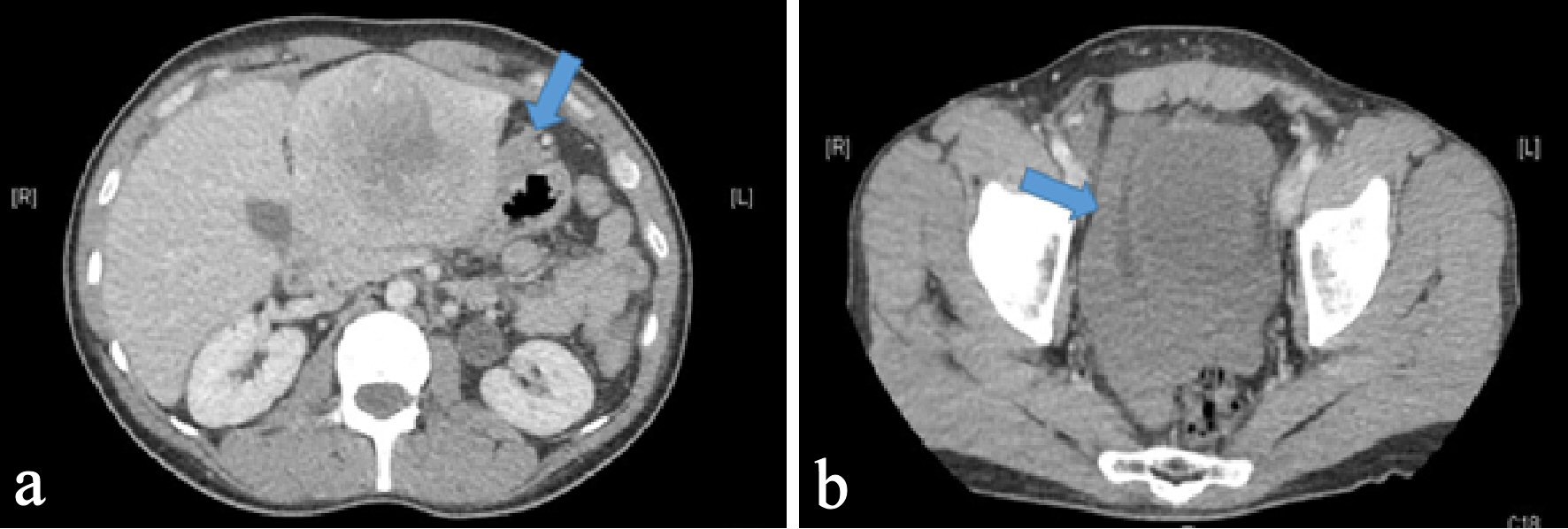
Figure 1. Axial section of CT abdomen and pelvis with intravenous contrast. (a) An 8 × 7 cm heterogeneous lobulated mass with central hypodensity in the left hepatic lobe with arrow pointing perilesional blood concerning for tumor rupture. (b) An arrow pointing hyperdensity fluid surrounding urinary bladder likely blood from tumor rupture.
Figure 2.
Figure 2. MRI of the abdomen with liver protocol post embolization showing a 7.7 × 6.6 cm well circumscribed expansile lesion in left lobe of liver with global hypo T2 signal intensity with areas of hyper T2 signal intensities as well as central stellate shape scar of low T2 signal intensity.
Figure 3.
Figure 3. Histopathologic examination of hematoxylin and eosin (H&E) stained liver biopsy specimen. (a) Tumor cells have hepatocellular appearance: large polygonal cells, ample eosinophilic cytoplasm, with focal bile pigment, marked nuclear atypia, and intranuclear pseudoinclusions. (b) Tumor cells are separated by hyaline collagen.