Gastroenterology Research, ISSN 1918-2805 print, 1918-2813 online, Open Access
Article copyright, the authors; Journal compilation copyright, Gastroenterol Res and Elmer Press Inc
Journal website http://www.gastrores.org

Case Report

Volume 11, Number 6, December 2018, pages 422-425


Isolated Gastric Metastasis of Endometrial Adenocarcinoma: First Case Report and Review of Pertinent Literature

Figures

Figure 1.
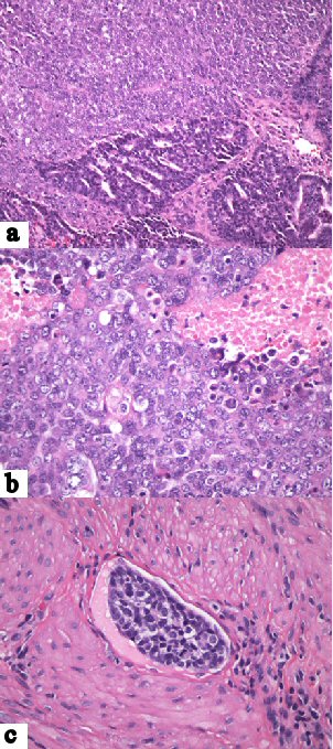
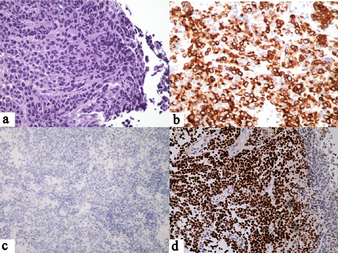
Figure 1. Gastric biopsy. (a) Large tumor cells with solid growth pattern, large, pleomorphic nuclei and high nuclear-to-cytoplasmic ratios. There are scattered intratumoral lymphocytes but no significant peritumoral lymphocytosis (H&E, × 40). (b) Positive tumor staining for CAM5.2 (× 20). (c) Negative tumor staining for CDX-2 (× 20). (d) Positive tumor staining for PAX-8 (× 20).
Figure 2.
Figure 2. Endometrial carcinoma. (a) Sharply demarcated regions of moderately and poorly differentiated carcinoma (H&E, × 20). (b) Poorly differentiated tumor component featuring solid growth pattern, large, pleomorphic nuclei, high nuclear-to-cytoplasmic ratios and necrosis (× 40). (c) Lymphovascular invasion by poorly differentiated component (× 40).