Gastroenterology Research, ISSN 1918-2805 print, 1918-2813 online, Open Access
Article copyright, the authors; Journal compilation copyright, Gastroenterol Res and Elmer Press Inc
Journal website http://www.gastrores.org

Case Report

Volume 11, Number 4, August 2018, pages 329-332


Spontaneous Expulsion per Rectum of a Colorectal Polyp: A Rare and Unusual Case

Figures

Figure 1.
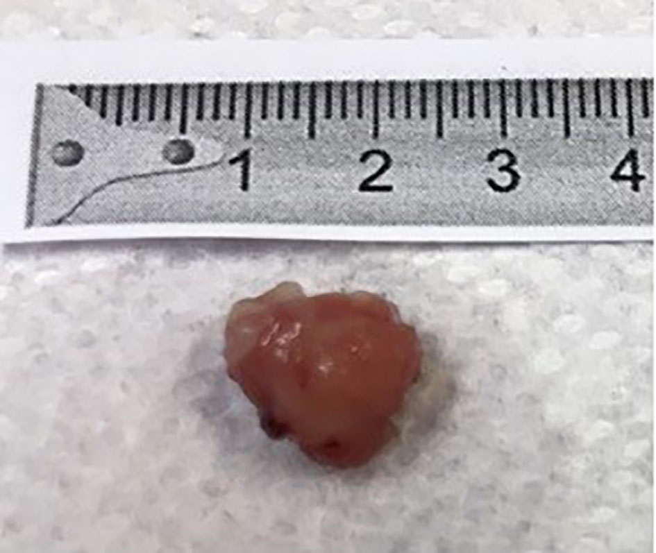
Figure 1. The specimen brought by the patient in a container after spontaneous expulsion per rectum. It appeared to be a colorectal polyp, roughly 1.4 cm in diameter.
Figure 2.
Figure 2. Histopathologic image of the colorectal polyp. The neoplastic glands are lined by hyperchromatic glandular epithelium. The elongated nuclei and reduced cytoplasmic mucin production are consistent with a large tubular adenoma. H&E stain. Low-power magnification.
Figure 3.
Figure 3. Endoscopic images of (a) a 1.8 cm pedunculated polyp in the descending colon, (b) an 8 - 9 mm pedunculated polyp in the distal transverse colon, (c) a 2 cm pedunculated polyp in the proximal transverse colon, and (d) a 2 cm sessile polyp in the ascending colon.